- Record: found
- Abstract: found
- Article: found
Initial Physician Experience with [ 18F]Flutemetamol Amyloid PET Imaging Following Availability for Routine Clinical Use in Japan
Read this article at
Abstract
Background:
Brain amyloid is a neuropathological hallmark of Alzheimer’s disease (AD). By visualizing brain amyloid, positron emission tomography (PET) may influence the diagnostic assessment and management of patients with cognitive impairment.
Objective:
As part of a Japanese post-approval study to measure the safety of [ 18F]flutemetamol PET, the association of amyloid PET results with changes in diagnosis and diagnostic confidence was assessed.
Methods:
Fifty-seven subjects were imaged for amyloid PET using [ 18F]flutemetamol at a single Japanese memory clinic. The cognitive diagnosis and referring physician’s confidence in the diagnosis were recorded before and after availability of PET results. Imaging started approximately 90 minutes after [ 18F]flutemetamol administration with approximately 185 MBq injected. PET images were acquired for 30 minutes.
Results:
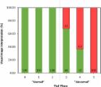
Amyloid PET imaging led to change in diagnosis in 15/44 clinical subjects (34%). Mean diagnostic confidence increased by approximately 20%, from 73% pre-scan to 93% post-scan, and this rise was fairly consistent across the main patient subgroups (mild cognitive impairment, AD, and non-AD) irrespective of the pre-scan diagnosis and scan result.
Conclusion:
The study examined the utility of amyloid PET imaging in a Japanese clinical cohort and highlighted the use of an etiological diagnosis in the presence of the amyloid scan. [ 18F]Flutemetamol PET led to a change in diagnosis in over 30% of cases and to an increase in diagnostic confidence by approximately 20% consistent with other reports.
Related collections
Most cited references16
- Record: found
- Abstract: found
- Article: found
Association of Amyloid Positron Emission Tomography With Changes in Diagnosis and Patient Treatment in an Unselected Memory Clinic Cohort: The ABIDE Project
- Record: found
- Abstract: found
- Article: not found