35
views
0
recommends
+1 Recommend
0 collections
    0
    shares
      • Record: found
      • Abstract: found
      • Article: found
      Is Open Access

      Update on Vascular Cognitive Impairment Associated with Subcortical Small-Vessel Disease

      review-article

      Read this article at

      Bookmark
          There is no author summary for this article yet. Authors can add summaries to their articles on ScienceOpen to make them more accessible to a non-specialist audience.

          Abstract

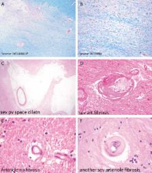
          Subcortical small-vessel disease (SSVD) is a disorder well characterized from the clinical, imaging, and neuropathological viewpoints. SSVD is considered the most prevalent ischemic brain disorder, increasing in frequency with age. Vascular risk factors include hypertension, diabetes, hyperlipidemia, elevated homocysteine, and obstructive sleep apnea. Ischemic white matter lesions are the hallmark of SSVD; other pathological lesions include arteriolosclerosis, dilatation of perivascular spaces, venous collagenosis, cerebral amyloid angiopathy, microbleeds, microinfarcts, lacunes, and large infarcts. The pathogenesis of SSVD is incompletely understood but includes endothelial changes and blood-brain barrier alterations involving metalloproteinases, vascular endothelial growth factors, angiotensin II, mindin/spondin, and the mammalian target of rapamycin pathway. Metabolic and genetic conditions may also play a role but hitherto there are few conclusive studies. Clinical diagnosis of SSVD includes early executive dysfunction manifested by impaired capacity to use complex information, to formulate strategies, and to exercise self-control. In comparison with Alzheimer’s disease (AD), patients with SSVD show less pronounced episodic memory deficits. Brain imaging has advanced substantially the diagnostic tools for SSVD. With the exception of cortical microinfarcts, all other lesions are well visualized with MRI. Diagnostic biomarkers that separate AD from SSVD include reduction of cerebrospinal fluid amyloid-β (Aβ) 42 and of the ratio Aβ 42/Aβ 40 often with increased total tau levels. However, better markers of small-vessel function of intracerebral blood vessels are needed. The treatment of SSVD remains unsatisfactory other than control of vascular risk factors. There is an urgent need of finding targets to slow down and potentially halt the progression of this prevalent, but often unrecognized, disorder.

          Related collections

          Most cited references188

          • Record: found
          • Abstract: found
          • Article: not found

          Gene dose of apolipoprotein E type 4 allele and the risk of Alzheimer's disease in late onset families.

          The apolipoprotein E type 4 allele (APOE-epsilon 4) is genetically associated with the common late onset familial and sporadic forms of Alzheimer's disease (AD). Risk for AD increased from 20% to 90% and mean age at onset decreased from 84 to 68 years with increasing number of APOE-epsilon 4 alleles in 42 families with late onset AD. Thus APOE-epsilon 4 gene dose is a major risk factor for late onset AD and, in these families, homozygosity for APOE-epsilon 4 was virtually sufficient to cause AD by age 80.
            Bookmark
            • Record: found
            • Abstract: found
            • Article: not found

            The role of apolipoprotein E in Alzheimer's disease.

            The epsilon4 allele of apolipoprotein E (APOE) is the major genetic risk factor for Alzheimer's disease (AD). Although there have been numerous studies attempting to elucidate the underlying mechanism for this increased risk, how apoE4 influences AD onset and progression has yet to be proven. However, prevailing evidence suggests that the differential effects of apoE isoforms on Abeta aggregation and clearance play the major role in AD pathogenesis. Other potential mechanisms, such as the differential modulation of neurotoxicity and tau phosphorylation by apoE isoforms as well as its role in synaptic plasticity and neuroinflammation, have not been ruled out. Inconsistent results among studies have made it difficult to define whether the APOE epsilon4 allele represents a gain of toxic function, a loss of neuroprotective function, or both. Therapeutic strategies based on apoE propose to reduce the toxic effects of apoE4 or to restore the physiological, protective functions of apoE.
              Bookmark
              • Record: found
              • Abstract: found
              • Article: not found

              The many faces of insulin-like peptide signalling in the brain.

              Central and peripheral insulin-like peptides (ILPs), which include insulin, insulin-like growth factor 1 (IGF1) and IGF2, exert many effects in the brain. Through their actions on brain growth and differentiation, ILPs contribute to building circuitries that subserve metabolic and behavioural adaptation to internal and external cues of energy availability. In the adult brain each ILP has distinct effects, but together their actions ultimately regulate energy homeostasis - they affect nutrient sensing and regulate neuronal plasticity to modulate adaptive behaviours involved in food seeking, including high-level cognitive operations such as spatial memory. In essence, the multifaceted activity of ILPs in the brain may be viewed as a system organization involved in the control of energy allocation.
                Bookmark

                Author and article information

                Journal
                J Alzheimers Dis
                J. Alzheimers Dis
                JAD
                Journal of Alzheimer's Disease
                IOS Press (Nieuwe Hemweg 6B, 1013 BG Amsterdam, The Netherlands )
                1387-2877
                1875-8908
                13 March 2018
                2018
                : 62
                : 3 , Special 20th Anniversary Issue
                : 1417-1441
                Affiliations
                [a ]Institute of Neuroscience and Physiology at Sahlgrenska Academy, University of Gothenburg , Gothenburg Sweden and Memory Clinic at Department of Neuropsychiatry, Sahlgrenska University, Hospital, Gothenburg, Sweden
                [b ]Department of Neurology, Methodist Neurological Institute , Houston, TX, USA
                [c ]Weill Cornell Medical College, Cornell University , New York, NY, USA
                [d ]Neuropathology Department, West Wing, John Radcliffe Hospital , Oxford, UK
                [e ]Nuffield Department of Clinical Neurosciences, University of Oxford , West Wing, John Radcliffe Hospital, Oxford, UK
                [f ]Institute of Medicine at Sahlgrenska Academy, University of Gothenburg , Gothenburg, Sweden
                [g ]1st Department of Neurology, Neurochemistry Unit, National and Kapodistrian University of Athens , Athens, Greece
                Author notes
                [1]

                These authors contributed equally to this work.

                [* ]Correspondence to: Anders Wallin, MD, PhD, Professor Institute of Neuroscience and Physiology at Sahlgrenska Academy, University of Gothenburg, Gothenburg, Sweden and Memory Clinic, Department of Neuropsychiatry, Sahlgrenska University Hospital, Gothenburg, Sweden. Tel.: +46 31 342 10 00; E-mail: anders.wallin@ 123456neuro.gu.se .
                Article
                JAD170803
                10.3233/JAD-170803
                5870030
                29562536
                696d7f31-4548-49f9-a4a5-5604cc980c0c
                © 2018 – IOS Press and the authors. All rights reserved

                This is an open access article distributed under the terms of the Creative Commons Attribution Non-Commercial (CC BY-NC 4.0) License, which permits unrestricted non-commercial use, distribution, and reproduction in any medium, provided the original work is properly cited.

                History
                : 15 November 2017
                Categories
                Review

                cerebral small vessel disease,classification,cerebrospinal fluid,diagnostic imaging,genetics,metabolism,pathology,pathophysiology,symptoms,cognitive impairment

                Comments

                Comment on this article