8
views
0
recommends
+1 Recommend
0 collections
    0
    shares
      • Record: found
      • Abstract: found
      • Article: not found

      Augmented Stat5 Signaling Bypasses Multiple Impediments to Lactogen-Mediated Proliferation in Human β-Cells

      research-article

      Read this article at

      ScienceOpenPublisherPMC
      Bookmark
          There is no author summary for this article yet. Authors can add summaries to their articles on ScienceOpen to make them more accessible to a non-specialist audience.

          Abstract

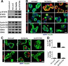
          Pregnancy in rodents is associated with a two- to threefold increase in β-cell mass, which is attributable to large increases in β-cell proliferation, complimented by increases in β-cell size, survival, and function and mediated mainly by the lactogenic hormones prolactin (PRL) and placental lactogens. In humans, however, β-cell mass does not increase as dramatically during pregnancy, and PRL fails to activate proliferation in human islets in vitro. To determine why, we explored the human PRL–prolactin receptor (hPRLR)–Janus kinase 2 (JAK2)–signal transducer and activator of transcription 5 (STAT5)–cyclin–cdk signaling cascade in human β-cells. Surprisingly, adult human β-cells express little or no PRLR. As expected, restoration of the hPRLR in human β-cells rescued JAK2-STAT5 signaling in response to PRL. However, rescuing hPRLR-STAT5 signaling nevertheless failed to confer proliferative ability on adult human β-cells in response to PRL. Surprisingly, mouse (but not human) Stat5a overexpression led to upregulation of cyclins D1–3 and cdk4, as well as their nuclear translocation, all of which are associated with β-cell cycle entry. Collectively, the findings show that human β-cells fail to proliferate in response to PRL for multiple reasons, one of which is a paucity of functional PRL receptors, and that murine Stat5 overexpression is able to bypass these impediments.

          Related collections

          Most cited references42

          • Record: found
          • Abstract: found
          • Article: not found

          Pancreatic beta-cell mass in European subjects with type 2 diabetes.

          Decreases in both beta-cell function and number can contribute to insulin deficiency in type 2 diabetes. Here, we quantified the beta-cell mass in pancreas obtained at autopsy of 57 type 2 diabetic (T2D) and 52 non-diabetic subjects of European origin. Sections from the body and tail were immunostained for insulin. The beta-cell mass was calculated from the volume density of beta-cells (measured by point-counting methods) and the weight of the pancreas. The pancreatic insulin concentration was measured in some of the subjects. beta-cell mass increased only slightly with body mass index (BMI). After matching for BMI, the beta-cell mass was 41% (BMI 15 years of overt diabetes respectively). Pancreatic insulin concentration was 30% lower in patients. In conclusion, the average beta-cell mass is about 39% lower in T2D subjects compared with matched controls. Its decrease with duration of the disease could be a consequence of diabetes that, with further impairment of insulin secretion, contributes to the progressive deterioration of glucose homeostasis. We do not believe that the small difference in beta-cell mass observed within 5 years of onset could cause diabetes in the absence of beta-cell dysfunction.
            Bookmark
            • Record: found
            • Abstract: found
            • Article: not found

            Serotonin Regulates Pancreatic β-Cell Mass during Pregnancy

            During pregnancy, the energy requirements of the fetus impose changes in maternal metabolism. Increasing insulin resistance in the mother maintains nutrient flow to the growing fetus, while prolactin and placental lactogen counterbalance this resistance and prevent maternal hyperglycemia by driving expansion of the maternal population of insulin-producing β-cells1–3. However, the exact mechanisms by which the lactogenic hormones drive β-cell expansion remain uncertain. Here we show that serotonin acts downstream of lactogen signaling to drive β-cell proliferation. Serotonin synthetic enzyme Tph1 and serotonin production increased sharply in β-cells during pregnancy or after treatment with lactogens in vitro. Inhibition of serotonin synthesis by dietary tryptophan restriction or Tph inhibition blocked β-cell expansion and induced glucose intolerance in pregnant mice without affecting insulin sensitivity. Expression of the Gαq-linked serotonin receptor Htr2b in maternal islets increased during pregnancy and normalized just prior to parturition, while expression of the Gαi-linked receptor Htr1d increased at the end of pregnancy and postpartum. Blocking Htr2b signaling in pregnant mice also blocked β-cell expansion and caused glucose intolerance. These studies reveal an integrated signaling pathway linking β-cell mass to anticipated insulin need during pregnancy. Modulators of this pathway, including medications and diet, may affect the risk of gestational diabetes4.
              Bookmark
              • Record: found
              • Abstract: found
              • Article: not found

              Adaptive changes in pancreatic beta cell fractional area and beta cell turnover in human pregnancy

              Aims/hypothesis We sought to establish the extent and basis for adaptive changes in beta cell numbers in human pregnancy. Methods Pancreas was obtained at autopsy from women who had died while pregnant (n = 18), post-partum (n = 6) or were not pregnant at or shortly before death (controls; n = 20). Pancreases were evaluated for fractional pancreatic beta cell area, islet size and islet fraction of beta cells, beta cell replication (Ki67) and apoptosis (TUNEL), and indirect markers of beta cell neogenesis (insulin-positive cells in ducts and scattered beta cells in pancreas). Results The pancreatic fractional beta cell area was increased by ∼1.4-fold in human pregnancy, with no change in mean beta cell size. In pregnancy there were more small islets rather than an increase in islet size or beta cells per islet. No increase in beta cell replication or change in beta cell apoptosis was detected, but duct cells positive for insulin and scattered beta cells were increased with pregnancy. Conclusions/interpretation The adaptive increase in beta cell numbers in human pregnancy is not as great as in most reports in rodents. This increase in humans is achieved by increased numbers of beta cells in apparently new small islets, rather than duplication of beta cells in existing islets, which is characteristic of pregnancy in rodents. Electronic supplementary material The online version of this article (doi:10.1007/s00125-010-1809-6) contains supplementary material, which is available to authorised users.
                Bookmark

                Author and article information

                Journal
                Diabetes
                Diabetes
                diabetes
                diabetes
                Diabetes
                Diabetes
                American Diabetes Association
                0012-1797
                1939-327X
                November 2015
                09 July 2015
                : 64
                : 11
                : 3784-3797
                Affiliations
                [1] 1Diabetes, Obesity and Metabolism Institute, Icahn School of Medicine at Mount Sinai, New York, NY
                [2] 2University of Maryland School of Medicine, Baltimore, MD
                [3] 3Duquesne University School of Nursing, Pittsburgh, PA
                [4] 4Department of Oncological Sciences, Icahn School of Medicine at Mount Sinai, New York, NY
                [5] 5Division of Endocrinology, Diabetes and Metabolism, University of Alabama at Birmingham, Birmingham, AL
                [6] 6Endocrinology Section, Birmingham VA Medical Center, Birmingham, AL
                Author notes
                Corresponding author: Hainan Chen, hainanchen2011@ 123456gmail.com , or Andrew F. Stewart, andrew.stewart@ 123456mssm.edu .
                Article
                0083
                10.2337/db15-0083
                4613973
                26159175
                6a4b16a9-2231-4a2a-b7fc-2993d1835c6c
                © 2015 by the American Diabetes Association. Readers may use this article as long as the work is properly cited, the use is educational and not for profit, and the work is not altered.
                History
                : 27 January 2015
                : 30 June 2015
                Page count
                Pages: 14
                Funding
                Funded by: Juvenile Diabetes Research Foundation http://dx.doi.org/10.13039/100000901
                Award ID: 1-2011-603
                Funded by: National Institutes of Health http://dx.doi.org/10.13039/100000002
                Award ID: R-01 DK 46395
                Award ID: U-01 DK089538
                Award ID: UC4 DK104211
                Funded by: James A. Haley Veterans’ Hospital http://dx.doi.org/10.13039/100006364
                Award ID: Merit Review (to S.J.F.)
                Categories
                Islet Studies

                Endocrinology & Diabetes
                Endocrinology & Diabetes

                Comments

                Comment on this article