23
views
0
recommends
+1 Recommend
0 collections
    0
    shares
      • Record: found
      • Abstract: found
      • Article: found
      Is Open Access

      Converting a Sulfenic Acid Reductase into a Disulfide Bond Isomerase

      research-article

      Read this article at

      Bookmark
          There is no author summary for this article yet. Authors can add summaries to their articles on ScienceOpen to make them more accessible to a non-specialist audience.

          Abstract

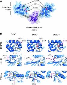
          Aims: Posttranslational formation of disulfide bonds is essential for the folding of many secreted proteins. Formation of disulfide bonds in a protein with more than two cysteines is inherently fraught with error and can result in incorrect disulfide bond pairing and, consequently, misfolded protein. Protein disulfide bond isomerases, such as DsbC of Escherichia coli, can recognize mis-oxidized proteins and shuffle the disulfide bonds of the substrate protein into their native folded state. Results: We have developed a simple blue/white screen that can detect disulfide bond isomerization in vivo, using a mutant alkaline phosphatase (PhoA*) in E. coli. We utilized this screen to isolate mutants of the sulfenic acid reductase (DsbG) that allowed this protein to act as a disulfide bond isomerase. Characterization of the isolated mutants in vivo and in vitro allowed us to identify key amino acid residues responsible for oxidoreductase properties of thioredoxin-like proteins such as DsbC or DsbG. Innovation and Conclusions: Using these key residues, we also identified and characterized interesting environmental homologs of DsbG with novel properties, thus demonstrating the capacity of this screen to discover and elucidate mechanistic details of in vivo disulfide bond isomerization. Antioxid. Redox Signal. 23, 945–957.

          Related collections

          Most cited references42

          • Record: found
          • Abstract: found
          • Article: not found

          Protein structure alignment by incremental combinatorial extension (CE) of the optimal path.

          A new algorithm is reported which builds an alignment between two protein structures. The algorithm involves a combinatorial extension (CE) of an alignment path defined by aligned fragment pairs (AFPs) rather than the more conventional techniques using dynamic programming and Monte Carlo optimization. AFPs, as the name suggests, are pairs of fragments, one from each protein, which confer structure similarity. AFPs are based on local geometry, rather than global features such as orientation of secondary structures and overall topology. Combinations of AFPs that represent possible continuous alignment paths are selectively extended or discarded thereby leading to a single optimal alignment. The algorithm is fast and accurate in finding an optimal structure alignment and hence suitable for database scanning and detailed analysis of large protein families. The method has been tested and compared with results from Dali and VAST using a representative sample of similar structures. Several new structural similarities not detected by these other methods are reported. Specific one-on-one alignments and searches against all structures as found in the Protein Data Bank (PDB) can be performed via the Web at http://cl.sdsc.edu/ce.html.
            Bookmark
            • Record: found
            • Abstract: found
            • Article: not found

            DNA microarray-mediated transcriptional profiling of the Escherichia coli response to hydrogen peroxide.

            The genome-wide transcription profile of Escherichia coli cells treated with hydrogen peroxide was examined with a DNA microarray composed of 4,169 E. coli open reading frames. By measuring gene expression in isogenic wild-type and oxyR deletion strains, we confirmed that the peroxide response regulator OxyR activates most of the highly hydrogen peroxide-inducible genes. The DNA microarray measurements allowed the identification of several new OxyR-activated genes, including the hemH heme biosynthetic gene; the six-gene suf operon, which may participate in Fe-S cluster assembly or repair; and four genes of unknown function. We also identified several genes, including uxuA, encoding mannonate hydrolase, whose expression might be repressed by OxyR, since their expression was elevated in the DeltaoxyR mutant strain. In addition, the induction of some genes was found to be OxyR independent, indicating the existence of other peroxide sensors and regulators in E. coli. For example, the isc operon, which specifies Fe-S cluster formation and repair activities, was induced by hydrogen peroxide in strains lacking either OxyR or the superoxide response regulators SoxRS. These results expand our understanding of the oxidative stress response and raise interesting questions regarding the nature of other regulators that modulate gene expression in response to hydrogen peroxide.
              Bookmark
              • Record: found
              • Abstract: found
              • Article: not found

              Structure, function, and mechanism of thioredoxin proteins.

              Thioredoxins are ubiquitous antioxidant enzymes that play important roles in many health-related cellular processes. As such, the fundamental knowledge of how these enzymes work is of prime importance for understanding cellular redox mechanisms and for laying the ground for the development of future therapeutic approaches. Over the past 40 years, a really impressive amount of data has been published on thioredoxins. Here, we review the most significant results that have contributed to our knowledge regarding the structure, the function, and the mechanism of these crucial enzymes.
                Bookmark

                Author and article information

                Journal
                Antioxid Redox Signal
                Antioxid. Redox Signal
                ars
                Antioxidants & Redox Signaling
                Mary Ann Liebert, Inc. (140 Huguenot Street, 3rd FloorNew Rochelle, NY 10801USA )
                1523-0864
                1557-7716
                20 October 2015
                20 October 2015
                : 23
                : 12
                : 945-957
                Affiliations
                [ 1 ]Protein Expression and Modification, New England Biolabs, Ipswich, Massachusetts.
                [ 2 ]Actelion, Allschwil, Switzerland.
                [ 3 ]Department of Microbiology and Immunobiology, Harvard Medical School , Boston, Massachusetts.
                [ 4 ]Novartis, Basel, Switzerland.
                [ 5 ]Howard Hughes Medical Institute Molecular, Cellular and Developmental Biology, University of Michigan , Ann Arbor, Michigan.
                Author notes
                [*]

                These authors contributed equally to this work.

                Address correspondence to: Dr. Mehmet Berkmen, Protein Expression and Modification, New England Biolabs, 240 County Road, Ipswich, MA 01938, E-mail: berkmen@ 123456neb.com
                Article
                10.1089/ars.2014.6235
                10.1089/ars.2014.6235
                4624244
                26191605
                6a8ce31a-c9d5-42d7-9657-3bc97da68ffb
                © Claire Chatelle et al 2015; Published by Mary Ann Liebert, Inc.

                This Open Access article is distributed under the terms of the Creative Commons Attribution Noncommercial License ( http://creativecommons.org/licenses/by-nc/4.0/) which permits any noncommercial use, distribution, and reproduction in any medium, provided the original author(s) and the source are credited.

                History
                : 22 December 2014
                : 28 April 2015
                : 13 May 2015
                Page count
                Figures: 4, References: 52, Pages: 13
                Categories
                Original Research Communications

                Comments

                Comment on this article