24
views
0
recommends
+1 Recommend
1 collections
    0
    shares

      International Journal of COPD (submit here)

      This international, peer-reviewed Open Access journal by Dove Medical Press focuses on pathophysiological processes underlying Chronic Obstructive Pulmonary Disease (COPD) interventions, patient focused education, and self-management protocols. Sign up for email alerts here.

      39,063 Monthly downloads/views I 2.893 Impact Factor I 5.2 CiteScore I 1.16 Source Normalized Impact per Paper (SNIP) I 0.804 Scimago Journal & Country Rank (SJR)

      • Record: found
      • Abstract: found
      • Article: found
      Is Open Access

      Fluticasone propionate and increased risk of pneumonia in COPD: is it PAFR-dependent?

      letter
      International Journal of Chronic Obstructive Pulmonary Disease
      Dove Medical Press

      Read this article at

      ScienceOpenPublisherPMC
      Bookmark
          There is no author summary for this article yet. Authors can add summaries to their articles on ScienceOpen to make them more accessible to a non-specialist audience.

          Abstract

          Dear editor It was with great interest that I read the recent comprehensive review by Christer Janson et al1 published in the International Journal of COPD, where authors discussed the possible mechanisms behind the increased risk of pneumonia in COPD patients using inhaled corticosteroids (ICSs), especially with fluticasone propionate (FP), where the risk was highest.1 It is an important area, and it is encouraging and reassuring that leading clinical journals are recognizing this. Understanding the fundamental mechanisms behind pneumococcal infections is critical.2 I would like to suggest that a broader discussion of new insights into the potential mechanisms contributing to the increased adherence of pneumococcus to airway wall and particularly in response to FP might has been appropriate with this opportunity. The prerequisite step for any pulmonary microbial infection is the adherence of pathogens to the respiratory mucosa via interaction between the host epithelial cells and the bacterial surface. A possible mechanism is through the interaction of phosphorylcholine, a molecular mimic of platelet-activating factor (PAF) present on the bacterial surface, while PAF receptor (PAFR) is expressed on the airway epithelium.3 Interestingly, both airway pathogens, pneumococcus and Haemophilus influenzae, adhere to and are engulfed by airway epithelial cells via the PAFR, thus evading the host immune responses and increasing their chances for colonization and infection.3 
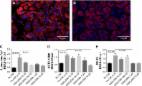
Our group previously published that PAFR expression increases in the airways of smokers and COPD patients but especially so in COPD.4 Importantly in this study, we also looked at the effects of FP on PAFR expression in COPD patients. Since ICSs increase the risk of pneumonia, we asked the question “Does ICS increase PAFR expression facilitating bacterial adhesion?” We surprisingly found that high doses of FP tend to increase PAFR expression in COPD patients. Overall, increase in epithelial PAFR expression was little, but it was evident that FP can upregulate PAFR expression – though FP certainly did not decrease it over 6 months.4 The intervention, though underpowered, still provided interesting inputs on the likely cause of observed vulnerability to pneumococcal infection in COPD patients treated with FP.4 We also observed an increased PAFR expression on small airway and alveolar type II pneumocytes and immune cells, suggesting pan airway PAFR expression in COPD.3,4 Further, our mechanistic in vitro infection model using immortalized lung epithelial cells demonstrated that a PAFR-specific chemical antagonist such as WEB-2086 significantly decreased the adherence and engulfment of H. influenzae and pneumococcus in a dose-dependent manner.5 These observations suggest that PAFR might be an important bacterial adhesion site, which is potentially upregulated in response to ICS treatment. Our findings in COPD might well be applicable to other chronic lung disease such as asthma and interstitial lung diseases.2 This recent paper by Christer Janson et al1 is a timely reminder that understanding of these mechanisms is of utmost importance and will stimulate further research. The main emphasis in the literature has been on bacterial colonizations in airways, but the mechanisms underlying initial bacterial epithelial adherence and consequent infections remain poorly understood.

          Most cited references7

          • Record: found
          • Abstract: found
          • Article: found
          Is Open Access

          Scientific rationale for the possible inhaled corticosteroid intraclass difference in the risk of pneumonia in COPD

          Inhaled corticosteroids (ICSs) treatment combined with long-acting β2-adrenoceptor agonists (LABAs) reduces the risk of exacerbations in COPD, but the use of ICSs is associated with increased incidence of pneumonia. There are indications that this association is stronger for fluticasone propionate than for budesonide. We have examined systematic reviews assessing the risk of pneumonia associated with fluticasone propionate and budesonide COPD therapy. Compared with placebo or LABAs, we found that fluticasone propionate was associated with 43%–78% increased risk of pneumonia, while only slightly increased risk or no risk was found for budesonide. We have evaluated conceivable mechanisms which may explain this difference and suggest that the higher pneumonia risk with fluticasone propionate treatment is caused by greater and more protracted immunosuppressive effects locally in the airways/lungs. These effects are due to the much slower dissolution of fluticasone propionate particles in airway luminal fluid, resulting in a slower uptake into the airway tissue and a much longer presence of fluticasone propionate in airway epithelial lining fluid.
            Bookmark
            • Record: found
            • Abstract: found
            • Article: found
            Is Open Access

            Budesonide and fluticasone propionate differentially affect the airway epithelial barrier

            Background COPD patients have a higher risk of pneumonia when treated with fluticasone propionate (FP) than with placebo, and a lower risk with budesonide (BUD). We hypothesized that BUD and FP differentially affect the mucosal barrier in response to viral infection and/or cigarette smoke. Methods We assessed protective effects of equivalent concentrations of BUD and FP on cytokine production and barrier function (electrical resistance) in human bronchial epithelial 16HBE cells and primary bronchial epithelial cells (PBECs) upon exposure to viral mimetic poly-(I:C) and/or cigarette smoke extract (CSE) or epidermal growth factor (EGF). Results BUD and FP were equally effective in suppressing poly-(I:C)- and/or CSE-induced IL-8 secretion in 16HBE and PBECs. Poly-(I:C) substantially decreased electrical resistance in 16HBE cells and both BUD and FP fully counteracted this effect. However, FP hardly affected 16HBE barrier dysfunction induced by CSE with/without poly-(I:C), whereas BUD (16 nM) provided full protection, an effect likely mediated by affecting EGFR-downstream target GSK-3β. Similarly, BUD, but not FP, significantly improved CSE-induced barrier dysfunction in PBECs. Finally, BUD, but not FP, exerted a modest but significant protective effect against Streptococcus Pneumoniae-induced barrier dysfunction, and BUD, but not FP, prevented cellular adhesion and/or internalization of these bacteria induced by poly-(I:C) in 16HBE. Conclusions Collectively, both BUD and FP efficiently control epithelial pro-inflammatory responses and barrier function upon mimicry of viral infection. Of potential clinical relevance, BUD more effectively counteracted CSE-induced barrier dysfunction, reinforcing the epithelial barrier and potentially limiting access of pathogens upon smoking in vivo.
              Bookmark
              • Record: found
              • Abstract: found
              • Article: found
              Is Open Access

              An antagonist of the platelet-activating factor receptor inhibits adherence of both nontypeable Haemophilus influenzae and Streptococcus pneumoniae to cultured human bronchial epithelial cells exposed to cigarette smoke

              Background COPD is emerging as the third largest cause of human mortality worldwide after heart disease and stroke. Tobacco smoking, the primary risk factor for the development of COPD, induces increased expression of platelet-activating factor receptor (PAFr) in the lung epithelium. Nontypeable Haemophilus influenzae (NTHi) and Streptococcus pneumoniae adhere to PAFr on the luminal surface of human respiratory tract epithelial cells. Objective To investigate PAFr as a potential drug target for the prevention of infections caused by the main bacterial drivers of acute exacerbations in COPD patients, NTHi and S. pneumoniae. Methods Human bronchial epithelial BEAS-2B cells were exposed to cigarette smoke extract (CSE). PAFr expression levels were determined using immunocytochemistry and quantitative polymerase chain reaction. The epithelial cells were challenged with either NTHi or S. pneumoniae labeled with fluorescein isothiocyanate, and bacterial adhesion was measured using immunofluorescence. The effect of a well-evaluated antagonist of PAFr, WEB-2086, on binding of the bacterial pathogens to BEAS-2B cells was then assessed. In silico studies of the tertiary structure of PAFr and the binding pocket for PAF and its antagonist WEB-2086 were undertaken. Results PAFr expression by bronchial epithelial cells was upregulated by CSE, and significantly associated with increased bacterial adhesion. WEB-2086 reduced the epithelial adhesion by both NTHi and S. pneumoniae to levels observed for non-CSE-exposed cells. Furthermore, it was nontoxic toward the bronchial epithelial cells. In silico analyses identified a binding pocket for PAF/WEB-2086 in the predicted PAFr structure. Conclusion WEB-2086 represents an innovative class of candidate drugs for inhibiting PAFr-dependent lung infections caused by the main bacterial drivers of smoking-related COPD.
                Bookmark

                Author and article information

                Journal
                Int J Chron Obstruct Pulmon Dis
                Int J Chron Obstruct Pulmon Dis
                International Journal of COPD
                International Journal of Chronic Obstructive Pulmonary Disease
                Dove Medical Press
                1176-9106
                1178-2005
                2017
                04 December 2017
                : 12
                : 3425-3427
                Affiliations
                Discipline of Laboratory Medicine, School of Health Sciences, Faculty of Health, University of Tasmania, Launceston, TAS, Australia
                [1 ]Respiratory, Allergy and Sleep Research Unit, Department of Medical Sciences, Uppsala University, Uppsala
                [2 ]Respiratory, Inflammation and Autoimmunity, AstraZeneca Nordic, Södertälje
                [3 ]Respiratory GMed, AstraZeneca Gothenburg, Mölndal, Sweden
                [4 ]Nottingham Respiratory Research Unit, City Hospital Campus, University of Nottingham, Nottingham, UK
                [5 ]Lung and Airway Research, National Institute of Environmental Medicine, Karolinska Institutet, Stockholm, Sweden
                Author notes
                Correspondence: Sukhwinder Singh Sohal, Discipline of Laboratory Medicine, School of Health Sciences, Faculty of Health, University of Tasmania, Locked Bag – 1322, Newnham Drive Launceston, Tasmania 7248, Australia, Tel +61 3 6324 5434, Email sssohal@ 123456utas.edu.au
                Correspondence: Christer Janson, Respiratory, Allergy and Sleep Research Unit, Department of Medical Sciences, Uppsala University, Uppsala, Sweden
                Article
                copd-12-3425
                10.2147/COPD.S154897
                5720348
                6bd36281-1304-4cfb-a12c-c6385f7abb31
                © 2017 Sohal. This work is published and licensed by Dove Medical Press Limited

                The full terms of this license are available at https://www.dovepress.com/terms.php and incorporate the Creative Commons Attribution – Non Commercial (unported, v3.0) License ( http://creativecommons.org/licenses/by-nc/3.0/). By accessing the work you hereby accept the Terms. Non-commercial uses of the work are permitted without any further permission from Dove Medical Press Limited, provided the work is properly attributed.

                History
                Categories
                Letter

                Respiratory medicine
                Respiratory medicine

                Comments

                Comment on this article