144
views
0
recommends
+1 Recommend
0 collections
    0
    shares
      • Record: found
      • Abstract: found
      • Article: not found

      Glucose Sensing in L Cells: A Primary Cell Study

      brief-report

      Read this article at

      ScienceOpenPublisherPMC
      Bookmark
          There is no author summary for this article yet. Authors can add summaries to their articles on ScienceOpen to make them more accessible to a non-specialist audience.

          Summary

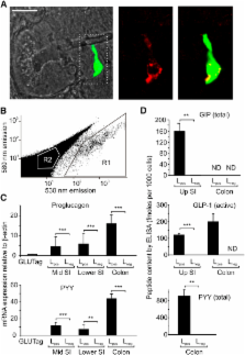
          Glucagon-like peptide-1 (GLP-1) is an enteric hormone that stimulates insulin secretion and improves glycaemia in type 2 diabetes. Although GLP-1-based treatments are clinically available, alternative strategies to increase endogenous GLP-1 release from L cells are hampered by our limited physiological understanding of this cell type. By generating transgenic mice with L cell-specific expression of a fluorescent protein, we studied the characteristics of primary L cells by electrophysiology, fluorescence calcium imaging, and expression analysis and show that single L cells are electrically excitable and glucose responsive. Sensitivity to tolbutamide and low-millimolar concentrations of glucose and α-methylglucopyranoside, assessed in single L cells and by hormone secretion from primary cultures, suggested that GLP-1 release is regulated by the activity of sodium glucose cotransporter 1 and ATP-sensitive K + channels, consistent with their high expression levels in purified L cells by quantitative RT-PCR. These and other pathways identified using this approach will provide exciting opportunities for future physiological and therapeutic exploration.

          Related collections

          Most cited references25

          • Record: found
          • Abstract: found
          • Article: not found

          Gut-expressed gustducin and taste receptors regulate secretion of glucagon-like peptide-1.

          Glucagon-like peptide-1 (GLP-1), released from gut endocrine L cells in response to glucose, regulates appetite, insulin secretion, and gut motility. How glucose given orally, but not systemically, induces GLP-1 secretion is unknown. We show that human duodenal L cells express sweet taste receptors, the taste G protein gustducin, and several other taste transduction elements. Mouse intestinal L cells also express alpha-gustducin. Ingestion of glucose by alpha-gustducin null mice revealed deficiencies in secretion of GLP-1 and the regulation of plasma insulin and glucose. Isolated small bowel and intestinal villi from alpha-gustducin null mice showed markedly defective GLP-1 secretion in response to glucose. The human L cell line NCI-H716 expresses alpha-gustducin, taste receptors, and several other taste signaling elements. GLP-1 release from NCI-H716 cells was promoted by sugars and the noncaloric sweetener sucralose, and blocked by the sweet receptor antagonist lactisole or siRNA for alpha-gustducin. We conclude that L cells of the gut "taste" glucose through the same mechanisms used by taste cells of the tongue. Modulating GLP-1 secretion in gut "taste cells" may provide an important treatment for obesity, diabetes and abnormal gut motility.
            Bookmark
            • Record: found
            • Abstract: found
            • Article: not found

            T1R3 and gustducin in gut sense sugars to regulate expression of Na+-glucose cotransporter 1.

            Dietary sugars are transported from the intestinal lumen into absorptive enterocytes by the sodium-dependent glucose transporter isoform 1 (SGLT1). Regulation of this protein is important for the provision of glucose to the body and avoidance of intestinal malabsorption. Although expression of SGLT1 is regulated by luminal monosaccharides, the luminal glucose sensor mediating this process was unknown. Here, we show that the sweet taste receptor subunit T1R3 and the taste G protein gustducin, expressed in enteroendocrine cells, underlie intestinal sugar sensing and regulation of SGLT1 mRNA and protein. Dietary sugar and artificial sweeteners increased SGLT1 mRNA and protein expression, and glucose absorptive capacity in wild-type mice, but not in knockout mice lacking T1R3 or alpha-gustducin. Artificial sweeteners, acting on sweet taste receptors expressed on enteroendocrine GLUTag cells, stimulated secretion of gut hormones implicated in SGLT1 up-regulation. Gut-expressed taste signaling elements involved in regulating SGLT1 expression could provide novel therapeutic targets for modulating the gut's capacity to absorb sugars, with implications for the prevention and/or treatment of malabsorption syndromes and diet-related disorders including diabetes and obesity.
              Bookmark
              • Record: found
              • Abstract: found
              • Article: not found

              Distribution of glucagon-like peptide-1 and other preproglucagon-derived peptides in the rat hypothalamus and brainstem.

              Central administration of the preproglucagon-derived peptide glucagon-like peptide-1 significantly inhibits ingestion of food and water, and glucagon-like peptide-1 binding sites are present in a multitude of central areas involved in the regulation of ingestional behaviour. To evaluate further the neuroanatomical organization of central glucagon-like peptide-1 containing neuronal circuits with potential implications on ingestional behaviour, we carried out a series of experiments in the rat demonstrating the topographical sites of synthesis and processing of the preproglucagon precursor followed by a chromatographic analysis of the processed fragments. In situ hybridization histochemistry revealed that preproglucagon encoding messenger RNA was expressed in a single population of neurons in the caudal portion of the nucleus of the solitary tract. Gel chromatographic analysis of hypothalamic and brainstem tissue extracts revealed that the preproglucagon precursor is processed in a fashion similar to that seen in the small intestine, preferentially giving rise to glicentin, glucagon-like peptide-1 and glucagon-like peptide-2. This single brain site of glucagon-like peptide-1 synthesis was subsequently confirmed by immunohistochemical demonstration of glucagon-like peptide-1-immunoreactive perikarya in the central and caudal parts of the nucleus of the solitary tract. Numerous sites containing glucagon-like peptide-1 immunoreactive fibres were, however, discovered in the forebrain including hypothalamic, thalamic and cortical areas. The densest innervation by glucagon-like peptide-1 immunoreactive nerve fibres was seen in the hypothalamic dorsomedial and paraventricular nuclei, but numerous glucagon-like peptide-1 immunoreactive fibres were also seen throughout the periventricular strata of the third ventricle. Dual-labelling immunohistochemistry for tyrosine hydroxylase and glucagon-like peptide-1 gave no evidence for co-localization of catecholamines and glucagon-like peptide-1 in neurons of the lower brainstem. To identify neurons of the nucleus of the solitary tract that project to the hypothalamic paraventricular nucleus, the retrograde tracer FluoroGold was injected into this hypothalamic target and dual immunocytochemical identification of glucagon-like peptide-1 and tyrosine hydroxylase-positive neurons was performed on brainstem sections containing retrogradely labelled perikarya. From this experiment it was seen that many of the retrogradely labelled neurons in the central portion of the nucleus of the solitary tract are catecholaminergic, while none is glucagon-like peptide-1 immunoreactive. In contrast, most of the retrogradely labelled neurons of the caudal portion of the nucleus of the solitary tract contain glucagon-like peptide-1. These observations further substantiate that glucagon-like peptide-1 neurons of the solitary tract constitute a distinct non-catecholaminergic cell group which projects to many targets, one of which is the hypothalamic paraventricular nucleus.
                Bookmark

                Author and article information

                Journal
                Cell Metab
                Cell Metabolism
                Cell Press
                1550-4131
                1932-7420
                06 December 2008
                06 December 2008
                : 8
                : 6-3
                : 532-539
                Affiliations
                [1 ]Cambridge Institute for Medical Research and Department of Clinical Biochemistry, Addenbrooke's Hospital, Hills Road, Cambridge CB2 0XY, UK
                Author notes
                []Corresponding author fmg23@ 123456cam.ac.uk
                [2]

                These authors contributed equally to this work

                Article
                CMET525
                10.1016/j.cmet.2008.11.002
                2697331
                19041768
                6bd5713b-5217-4932-a3d8-4d7ee315c9b5
                © 2008 ELL & Excerpta Medica.

                This document may be redistributed and reused, subject to certain conditions.

                History
                : 2 May 2008
                : 28 August 2008
                : 11 November 2008
                Categories
                Short Article

                Cell biology
                humdisease
                Cell biology
                humdisease

                Comments

                Comment on this article