16
views
0
recommends
+1 Recommend
0 collections
    0
    shares
      • Record: found
      • Abstract: found
      • Article: not found

      Touch and Tactile Neuropathic Pain Sensitivity Are Set by Corticospinal Projections

      research-article

      Read this article at

      ScienceOpenPublisherPMC
      Bookmark
          There is no author summary for this article yet. Authors can add summaries to their articles on ScienceOpen to make them more accessible to a non-specialist audience.

          Abstract

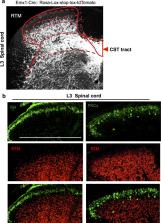
          Current models of somatosensory perception emphasize transmission from primary sensory neurons to the spinal cord and on to the brain 14 . Mental influence on perception is largely assumed to be acting locally within the brain. We have now examined if there is top-down control of sensory inflow through the spinal cord directly by the cortex. Although traditionally viewed as a primary motor pathway 5 , a subset of corticospinal neurons (CSNs) originating in the S1/S2 somatosensory cortex directly innervate the spinal dorsal horn via corticospinal tract (CST) axons. We show here that either reduction in somatosensory CSN activity or transection of the CST selectively impairs behavioral responses to light touch without altering responses to noxious stimuli. Moreover, such CSN manipulation greatly attenuates tactile allodynia in a peripheral neuropathic pain model. Tactile stimulation activates somatosensory CSNs and their corticospinal projections facilitate light touch-evoked activity of cholecystokinin (CCK +) interneurons in the deep dorsal horn. This represents a touch-driven feed forward spinal-cortical-spinal sensitization loop, which is important for the recruitment of spinal nociceptive neurons under tactile allodynia. These results reveal direct cortical modulation of normal and pathological tactile sensory processing in the spinal cord and open up opportunities for new treatments for neuropathic pain.

          Related collections

          Most cited references27

          • Record: found
          • Abstract: found
          • Article: not found

          A resource of Cre driver lines for genetic targeting of GABAergic neurons in cerebral cortex.

          A key obstacle to understanding neural circuits in the cerebral cortex is that of unraveling the diversity of GABAergic interneurons. This diversity poses general questions for neural circuit analysis: how are these interneuron cell types generated and assembled into stereotyped local circuits and how do they differentially contribute to circuit operations that underlie cortical functions ranging from perception to cognition? Using genetic engineering in mice, we have generated and characterized approximately 20 Cre and inducible CreER knockin driver lines that reliably target major classes and lineages of GABAergic neurons. More select populations are captured by intersection of Cre and Flp drivers. Genetic targeting allows reliable identification, monitoring, and manipulation of cortical GABAergic neurons, thereby enabling a systematic and comprehensive analysis from cell fate specification, migration, and connectivity, to their functions in network dynamics and behavior. As such, this approach will accelerate the study of GABAergic circuits throughout the mammalian brain. Copyright © 2011 Elsevier Inc. All rights reserved.
            Bookmark
            • Record: found
            • Abstract: found
            • Article: not found

            Spared nerve injury: an animal model of persistent peripheral neuropathic pain.

            Peripheral neuropathic pain is produced by multiple etiological factors that initiate a number of diverse mechanisms operating at different sites and at different times and expressed both within, and across different disease states. Unraveling the mechanisms involved requires laboratory animal models that replicate as far as possible, the different pathophysiological changes present in patients. It is unlikely that a single animal model will include the full range of neuropathic pain mechanisms. A feature of several animal models of peripheral neuropathic pain is partial denervation. In the most frequently used models a mixture of intact and injured fibers is created by loose ligation of either the whole (Bennett GJ, Xie YK. A peripheral mononeuropathy in rat that produces disorders of pain sensation like those seen in man. Pain 1988;33:87-107) or a tight ligation of a part (Seltzer Z, Dubner R, Shir Y. A novel behavioral model of neuropathic pain disorders produced in rats by partial sciatic nerve injury. Pain 1990;43:205-218) of a large peripheral nerve, or a tight ligation of an entire spinal segmental nerve (Kim SH, Chung JM. An experimental model for peripheral neuropathy produced by segmental spinal nerve ligation in the rat. Pain 1992;50:355-363). We have developed a variant of partial denervation, the spared nerve injury model. This involves a lesion of two of the three terminal branches of the sciatic nerve (tibial and common peroneal nerves) leaving the remaining sural nerve intact. The spared nerve injury model differs from the Chung spinal segmental nerve, the Bennett chronic constriction injury and the Seltzer partial sciatic nerve injury models in that the co-mingling of distal intact axons with degenerating axons is restricted, and it permits behavioral testing of the non-injured skin territories adjacent to the denervated areas. The spared nerve injury model results in early ( 6 months), robust (all animals are responders) behavioral modifications. The mechanical (von Frey and pinprick) sensitivity and thermal (hot and cold) responsiveness is increased in the ipsilateral sural and to a lesser extent saphenous territories, without any change in heat thermal thresholds. Crush injury of the tibial and common peroneal nerves produce similar early changes, which return, however to baseline at 7-9 weeks. The spared nerve injury model may provide, therefore, an additional resource for unraveling the mechanisms responsible for the production of neuropathic pain.
              Bookmark
              • Record: found
              • Abstract: found
              • Article: not found

              Long-term dynamics of CA1 hippocampal place codes

              Via Ca2+-imaging in freely behaving mice that repeatedly explored a familiar environment, we tracked thousands of CA1 pyramidal cells' place fields over weeks. Place coding was dynamic, for each day the ensemble representation of this environment involved a unique subset of cells. Yet, cells within the ∼15–25% overlap between any two of these subsets retained the same place fields, which sufficed to preserve an accurate spatial representation across weeks.
                Bookmark

                Author and article information

                Journal
                0410462
                6011
                Nature
                Nature
                Nature
                0028-0836
                1476-4687
                6 August 2018
                12 September 2018
                September 2018
                12 March 2019
                : 561
                : 7724
                : 547-550
                Affiliations
                [1 ]F.M. Kirby Neurobiology Center and Department of Neurology, Boston Children’s Hospital, 300 Longwood Avenue, Boston, MA 02115, USA
                [2 ]Departments of Neurology, Harvard Medical School, 300 Longwood Avenue, Boston, MA 02115, USA
                [3 ]Neurosurgery Department, Johns Hopkins School of Medicine, Baltimore, MD21205, USA
                [4 ]Unit on Neural Circuits and Adaptive Behaviors, Clinical and Translational Neuroscience Branch, National Institute of Mental Health, National Institutes of Health, Bethesda, MD 20892, USA
                [5 ]Department of Neurology and Research Center of Neurology in Second Affiliated Hospital, Key Laboratory of Medical Neurobiology of Zhejiang Province, Zhejiang University School of Medicine, Hangzhou, China
                [6 ]Department of Child Health Care, Children’s Hospital of Nanjing Medical University, Nanjing, 210008, China
                [7 ]Beth Israel Deaconess Medical Center, 330 Brookline Avenue, Boston, MA 02215, USA
                [8 ]Departments of Neurobiology, Harvard Medical School, 300 Longwood Avenue, Boston, MA 02115, USA
                Author notes
                [* ]Correspondence and requests for materials should be addressed to Z.H. ( Zhigang.he@ 123456childrens.harvard.edu ), C.J.W. ( clifford.woolf@ 123456childrens.harvard.edu ), or K.H.W. ( wkuan@ 123456mail.nih.gov )
                [9]

                These authors contributed equally to this work.

                Article
                NIHMS1500757
                10.1038/s41586-018-0515-2
                6163083
                30209395
                6dd39cf5-13c4-4ac2-a45a-9e0faeb6d40b

                Users may view, print, copy, and download text and data-mine the content in such documents, for the purposes of academic research, subject always to the full Conditions of use: http://www.nature.com/authors/editorial_policies/license.html#terms

                Reprints and permissions information is available at www.nature.com/reprints.

                History
                Categories
                Article

                Uncategorized
                Uncategorized

                Comments

                Comment on this article