14
views
0
recommends
+1 Recommend
0 collections
    0
    shares
      • Record: found
      • Abstract: found
      • Article: found
      Is Open Access

      Efficacy of rituximab in non-paraneoplastic autoimmune retinopathy

      research-article

      Read this article at

      Bookmark
          There is no author summary for this article yet. Authors can add summaries to their articles on ScienceOpen to make them more accessible to a non-specialist audience.

          Abstract

          Background

          Autoimmune retinopathy (AIR) is a rare but potentially blinding condition that is often underdiagnosed. Common features in AIR presentation include rapidly progressive vision loss with abnormal electrophysiological responses of the retina associated with positive anti-retinal antibodies. AIR is also challenging to treat, and thus, the introduction of new potential therapeutic agents is welcomed. The goal of this communication is to assess the effects of rituximab infusions on electroretinogram (ERG) responses and visual function outcomes in patients with non-paraneoplastic autoimmune retinopathy (npAIR).

          Results

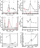
          Following infusion(s), three out of five patients showed no evidence of disease progression or improved, while two patients continued to progress on ERG. One patient demonstrated improvement in visual acuity (2 lines) in both eyes. ERG responses provided objective monitoring of patients’ visual function and response to immunosuppression over time.

          Conclusions

          These findings suggest that patients with npAIR unresponsive to other immunosuppression therapies may benefit from rituximab infusion, although stabilization rather than improvement was more frequently the outcome in our case series. Furthermore, regularly scheduled ERG follow-up examinations are recommended for monitoring patients’ progression during treatment.

          Related collections

          Most cited references45

          • Record: found
          • Abstract: found
          • Article: not found

          B cell development pathways.

          B cell development is a highly regulated process whereby functional peripheral subsets are produced from hematopoietic stem cells, in the fetal liver before birth and in the bone marrow afterward. Here we review progress in understanding some aspects of this process in the mouse bone marrow, focusing on delineation of the earliest stages of commitment, on pre-B cell receptor selection, and B cell tolerance during the immature-to-mature B cell transition. Then we note some of the distinctions in hematopoiesis and pre-B selection between fetal liver and adult bone marrow, drawing a connection from fetal development to B-1/CD5(+) B cells. Finally, focusing on CD5(+) cells, we consider the forces that influence the generation and maintenance of this distinctive peripheral B cell population, enriched for natural autoreactive specificities that are encoded by particular germline V(H)-V(L) combinations.
            Bookmark
            • Record: found
            • Abstract: found
            • Article: found
            Is Open Access

            Antibody to aquaporin-4 in the long-term course of neuromyelitis optica

            Neuromyelitis optica (NMO) is a severe inflammatory CNS disorder of putative autoimmune aetiology, which predominantly affects the spinal cord and optic nerves. Recently, a highly specific serum reactivity to CNS microvessels, subpia and Virchow–Robin spaces was described in patients with NMO [called NMO–IgG (NMO–immunoglobulin G)]. Subsequently, aquaporin-4 (AQP4), the most abundant water channel in the CNS, was identified as its target antigen. Strong support for a pathogenic role of the antibody would come from studies demonstrating a correlation between AQP4-Ab (AQP4-antibody) titres and the clinical course of disease. In this study, we determined AQP4-Ab serum levels in 96 samples from eight NMO–IgG positive patients (median follow-up 62 months) in a newly developed fluorescence-based immunoprecipitation assay employing recombinant human AQP4. We found that AQP4-Ab serum levels correlate with clinical disease activity, with relapses being preceded by an up to 3-fold increase in AQP4-Ab titres, which was not paralleled by a rise in other serum autoantibodies in one patient. Moreover, AQP4-Ab titres were found to correlate with CD19 cell counts during therapy with rituximab. Treatment with immunosuppressants such as rituximab, azathioprine and cyclophosphamide resulted in a marked reduction in antibody levels and relapse rates. Our results demonstrate a strong relationship between AQP4-Abs and clinical state, and support the hypothesis that these antibodies are involved in the pathogenesis of NMO.
              Bookmark
              • Record: found
              • Abstract: found
              • Article: not found

              Autoantibodies against retinal proteins in paraneoplastic and autoimmune retinopathy

              Background Autoimmune retinal degeneration may occur in patients who present with sudden or, less commonly, subacute loss of vision of retinal origin, associated with an abnormal ERG, through the action of autoantibodies against retinal proteins. Often the patients are initially diagnosed with or suspected of having a paraneoplastic retinopathy (PR), such as cancer-associated retinopathy (CAR). However, there is limited information on the occurrence, the specificity of autoantibodies in these patients, and their association with clinical symptoms. Methods Sera were obtained from 193 retinopathy patients who presented with clinical symptoms resembling PR or autoimmune retinopathy (AR), including sudden painless loss of vision, typically associated with visual field defects and photopsias, and abnormal rod and/or cone responses on the electroretinogram (ERG). Sera were tested for the presence of anti-retinal autoantibodies by Western blot analysis using proteins extracted from human retina and by immunohistochemistry. Autoantibody titers against recoverin and enolase were measured by ELISA. Results We identified a higher prevalence of anti-retinal autoantibodies in retinopathy patients. Ninety-one patients' sera (47.1%) showed autoantibodies of various specificities with a higher incidence of antibodies present in retinopathy patients diagnosed with cancer (33/52; 63.5%; p = 0.009) than in retinopathy patients without cancer (58/141; 41.1%). The average age of PR patients was 62.0 years, and that of AR patients was 55.9 years. Autoantibodies against recoverin (p23) were only present in the sera of PR patients, autoantibodies against unknown p35 were more common in patients with AR, while anti-enolase (anti-p46) autoantibodies were nearly equally distributed in the sera of patients with PR and those with AR. In the seropositive patients, the autoantibodies persisted over a long period of time – from months to years. A rebound in anti-recoverin autoantibody titer was found to be associated with exacerbations in visual symptoms but not in the recurrence of cancer. When compared to sera from healthy subjects, autoantibodies against retinal proteins from both groups of patients were cytotoxic to retinal cells, indicating their pathogenic potential. Conclusions These studies showed that patients with sudden or subacute, unexplained loss of vision of retinal origin have anti-retinal antibodies in a broad range of specificity and indicate the need for autoantibody screening. Follow-up tests of antibody levels may be useful as a biomarker of disease activity associated with worsening of vision. Moreover, the heterogeneity in autoantibody specificity may explain the variation and complexity of clinical symptoms in retinopathy patients.
                Bookmark

                Author and article information

                Contributors
                kateboudro@gmail.com
                sj2750@columbia.edu
                js3899@cumc.columbia.edu
                kaspar.schuerch@bluewin.ch
                wl2355@cumc.columbia.edu
                thiagogeorge@hotmail.com
                212-342-1189 , phosphodiesterase.gamma@gmail.com
                Journal
                Orphanet J Rare Dis
                Orphanet J Rare Dis
                Orphanet Journal of Rare Diseases
                BioMed Central (London )
                1750-1172
                15 July 2017
                15 July 2017
                2017
                : 12
                : 129
                Affiliations
                [1 ]ISNI 0000 0001 2292 3357, GRID grid.14848.31, Department of Ophthalmology, , University of Montreal, ; Montreal, Canada
                [2 ]ISNI 0000 0001 2285 2675, GRID grid.239585.0, Jonas Children’s Vision Care, and Bernard & Shirlee Brown Glaucoma Laboratory, , Columbia University Medical Center, ; New York, NY USA
                [3 ]ISNI 0000000419368729, GRID grid.21729.3f, Department of Ophthalmology, , Columbia University, ; New York, NY USA
                [4 ]ISNI 0000 0001 0693 2202, GRID grid.262863.b, , State University of New York Downstate Medical Center, ; Brooklyn, NY USA
                [5 ]ISNI 0000 0001 0514 7202, GRID grid.411249.b, Department of Ophthalmology, , Federal University of Sao Paulo (UNIFESP), ; São Paulo, Brazil
                [6 ]ISNI 0000 0001 2167 4168, GRID grid.412371.2, Department of Ophthalmology, , Federal University of Espírito Santo (UFES), ; Vitória, Brazil
                [7 ]ISNI 0000000419368729, GRID grid.21729.3f, Department of Pathology & Cell Biology, Institute of Human Nutrition, , College of Physicians and Surgeons, Columbia University, ; New York, NY USA
                Article
                680
                10.1186/s13023-017-0680-7
                5512938
                28709429
                702c0405-d9e9-47a4-9221-10fb876c6a9c
                © The Author(s). 2017

                Open AccessThis article is distributed under the terms of the Creative Commons Attribution 4.0 International License ( http://creativecommons.org/licenses/by/4.0/), which permits unrestricted use, distribution, and reproduction in any medium, provided you give appropriate credit to the original author(s) and the source, provide a link to the Creative Commons license, and indicate if changes were made. The Creative Commons Public Domain Dedication waiver ( http://creativecommons.org/publicdomain/zero/1.0/) applies to the data made available in this article, unless otherwise stated.

                History
                : 23 April 2017
                : 5 July 2017
                Funding
                Funded by: FundRef http://dx.doi.org/10.13039/100000002, National Institutes of Health;
                Award ID: 5P30EY019007
                Award ID: R01EY018213
                Award ID: R01EY024698
                Award ID: R01EY026682
                Award ID: R21AG050437
                Award Recipient :
                Categories
                Research
                Custom metadata
                © The Author(s) 2017

                Infectious disease & Microbiology
                autoimmune retinopathy,rituximab,treatment,multi-modal imaging,electroretinography

                Comments

                Comment on this article