19
views
0
recommends
+1 Recommend
0 collections
    0
    shares
      • Record: found
      • Abstract: found
      • Article: found
      Is Open Access

      Cognitive deficits and Alzheimer-like neuropathological impairments during adolescence in a rat model of type 2 diabetes mellitus

      research-article

      Read this article at

      Bookmark
          There is no author summary for this article yet. Authors can add summaries to their articles on ScienceOpen to make them more accessible to a non-specialist audience.

          Abstract

          Abstract

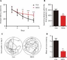
          Numerous studies have shown that many patients who suffer from type 2 diabetes mellitus exhibit cognitive dysfunction and neuronal synaptic impairments. Therefore, growing evidence suggests that type 2 diabetes mellitus has a close relationship with occurrence and progression of neurodegeneration and neural impairment in Alzheimer's disease. However, the relationship between metabolic disorders caused by type 2 diabetes mellitus and neurodegeneration and neural impairments in Alzheimer's disease is still not fully determined. Thus, in this study, we replicated a type 2 diabetic animal model by subcutaneous injection of newborn Sprague-Dawley rats with monosodium glutamate during the neonatal period. At 3 months old, the Barnes maze assay was performed to evaluate spatial memory function. Microelectrodes were used to measure electrophysiological function in the hippocampal CA1 region. Western blot assay was used to determine expression levels of glutamate ionotropic receptor NMDA type subunit 2A (GluN2A) and GluN2B in the hippocampus. Enzyme-linked immunosorbent assay was used to determine levels of interleukin-1β, tumor necrosis factor α, and interleukin-6 in the hippocampus and cerebral cortex, as well as hippocampal amyloid beta (Aβ) 1–40 and Aβ 1–42 levels. Our results showed that in the rat model of type 2 diabetes mellitus caused by monosodium glutamate exposure during the neonatal period, latency was prolonged and the number of errors increased in the Barnes maze. Further, latency was increased and time in the escape platform quadrant shortened. Number of times crossing the platform was also reduced in the Morris water maze. After high frequency stimulation of the hippocampus, synaptic transmission was inhibited, expression of GluN2A and GluN2B were decreased in the hippocampus, expression of interleukin 1β, interleukin 6, and tumor necrosis factor α was increased in the hippocampus and cortex, and levels of Aβ 1–40 and Aβ 1–42 were increased in the hippocampus. These findings confirm that type 2 diabetes mellitus induced by neonatal monosodium glutamate exposure results in Alzheimer-like neuropathological changes and further causes cognitive deficits and neurodegeneration in young adulthood.

          Related collections

          Most cited references55

          • Record: found
          • Abstract: found
          • Article: not found

          Inflammation and Alzheimer's disease.

          Inflammation clearly occurs in pathologically vulnerable regions of the Alzheimer's disease (AD) brain, and it does so with the full complexity of local peripheral inflammatory responses. In the periphery, degenerating tissue and the deposition of highly insoluble abnormal materials are classical stimulants of inflammation. Likewise, in the AD brain damaged neurons and neurites and highly insoluble amyloid beta peptide deposits and neurofibrillary tangles provide obvious stimuli for inflammation. Because these stimuli are discrete, microlocalized, and present from early preclinical to terminal stages of AD, local upregulation of complement, cytokines, acute phase reactants, and other inflammatory mediators is also discrete, microlocalized, and chronic. Cumulated over many years, direct and bystander damage from AD inflammatory mechanisms is likely to significantly exacerbate the very pathogenic processes that gave rise to it. Thus, animal models and clinical studies, although still in their infancy, strongly suggest that AD inflammation significantly contributes to AD pathogenesis. By better understanding AD inflammatory and immunoregulatory processes, it should be possible to develop anti-inflammatory approaches that may not cure AD but will likely help slow the progression or delay the onset of this devastating disorder.
            Bookmark
            • Record: found
            • Abstract: found
            • Article: not found

            The relative contributions of insulin resistance and beta-cell dysfunction to the pathophysiology of Type 2 diabetes.

            S E Kahn (2003)
            The relative contributions of insulin resistance and beta-cell dysfunction to the pathophysiology of Type 2 diabetes have been debated extensively. The concept that a feedback loop governs the interaction of the insulin-sensitive tissues and the beta cell as well as the elucidation of the hyperbolic relationship between insulin sensitivity and insulin secretion explains why insulin-resistant subjects exhibit markedly increased insulin responses while those who are insulin-sensitive have low responses. Consideration of this hyperbolic relationship has helped identify the critical role of beta-cell dysfunction in the development of Type 2 diabetes and the demonstration of reduced beta-cell function in high risk subjects. Furthermore, assessments in a number of ethnic groups emphasise that beta-cell function is a major determinant of oral glucose tolerance in subjects with normal and reduced glucose tolerance and that in all populations the progression from normal to impaired glucose tolerance and subsequently to Type 2 diabetes is associated with declining insulin sensitivity and beta-cell function. The genetic and molecular basis for these reductions in insulin sensitivity and beta-cell function are not fully understood but it does seem that body-fat distribution and especially intra-abdominal fat are major determinants of insulin resistance while reductions in beta-cell mass contribute to beta-cell dysfunction. Based on our greater understanding of the relative roles of insulin resistance and beta-cell dysfunction in Type 2 diabetes, we can anticipate advances in the identification of genes contributing to the development of the disease as well as approaches to the treatment and prevention of Type 2 diabetes.
              Bookmark
              • Record: found
              • Abstract: found
              • Article: not found

              Long-term potentiation and memory.

              M A Lynch (2004)
              One of the most significant challenges in neuroscience is to identify the cellular and molecular processes that underlie learning and memory formation. The past decade has seen remarkable progress in understanding changes that accompany certain forms of acquisition and recall, particularly those forms which require activation of afferent pathways in the hippocampus. This progress can be attributed to a number of factors including well-characterized animal models, well-defined probes for analysis of cell signaling events and changes in gene transcription, and technology which has allowed gene knockout and overexpression in cells and animals. Of the several animal models used in identifying the changes which accompany plasticity in synaptic connections, long-term potentiation (LTP) has received most attention, and although it is not yet clear whether the changes that underlie maintenance of LTP also underlie memory consolidation, significant advances have been made in understanding cell signaling events that contribute to this form of synaptic plasticity. In this review, emphasis is focused on analysis of changes that occur after learning, especially spatial learning, and LTP and the value of assessing these changes in parallel is discussed. The effect of different stressors on spatial learning/memory and LTP is emphasized, and the review concludes with a brief analysis of the contribution of studies, in which transgenic animals were used, to the literature on memory/learning and LTP.
                Bookmark

                Author and article information

                Journal
                Neural Regen Res
                Neural Regen Res
                NRR
                Neural Regeneration Research
                Medknow Publications & Media Pvt Ltd (India )
                1673-5374
                1876-7958
                November 2018
                : 13
                : 11
                : 1995-2004
                Affiliations
                [1 ]Department of Pathophysiology, Henan Medical College, Zhengzhou, Henan Province, China
                [2 ]Henan Key Laboratory of Degenerative Brain Disease, Henan Medical College, Zhengzhou, Henan Province, China
                [3 ]Department of Pathophysiology, School of Basic Medicine and the Collaborative Innovation Center for Brain Science, Key Laboratory of Ministry of Education of China for Neurological Disorders, Tongji Medical College, Huazhong University of Science and Technology, Wuhan, Hubei Province, China
                [4 ]Department of Pathology, Wuhan Children's Hospital, Wuhan, Hubei Province, China
                Author notes
                [* ] Correspondence to: Li Wang, kyk02@ 123456126.com .

                Author contributions: LW and LJ designed this study. LJ and YPL prepared all figures and wrote the manuscript. YPL and LR established experimental animal models and conducted the blood sample assays. YPL, LR and GJB performed the behavioral tests. QF and LJ conducted data analysis. LJ performed the western blot assay. YPL conducted the ELISA assays. All authors approved the final version of the paper.

                [#]

                These authors contributed equally to this work.

                Author information
                http://orcid.org/0000-0001-6205-5881
                Article
                NRR-13-1995
                10.4103/1673-5374.239448
                6183048
                30233075
                70c94b10-d53a-4765-81ad-8a07c633bbbc
                Copyright: © Neural Regeneration Research

                This is an open access journal, and articles are distributed under the terms of the Creative Commons Attribution-NonCommercial-ShareAlike 4.0 License, which allows others to remix, tweak, and build upon the work non-commercially, as long as appropriate credit is given and the new creations are licensed under the identical terms.

                History
                : 04 July 2018
                Categories
                Research Article

                nerve regeneration,type 2 diabetes mellitus,alzheimer's disease,monosodium glutamate,neonatal period,cognitive deficits,hyperglycemia,hyperinsulinemia,insulin resistance,neural regeneration

                Comments

                Comment on this article