46
views
0
recommends
+1 Recommend
0 collections
    0
    shares
      • Record: found
      • Abstract: found
      • Article: not found

      New single-molecule speckle microscopy reveals modification of the retrograde actin flow by focal adhesions at nanometer scales

      research-article

      Read this article at

      ScienceOpenPublisherPMC
      Bookmark
          There is no author summary for this article yet. Authors can add summaries to their articles on ScienceOpen to make them more accessible to a non-specialist audience.

          Abstract

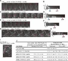
          This paper introduces a new, easy-to-use method of fluorescence single-molecule speckle microscopy for actin with nanometer-scale accuracy. This new method reveals that actin flows in front of mature focal adhesions (FAs) are fast and biased toward FAs, suggesting that mature FAs are actively engaged in pulling and remodeling the local actin network.

          Abstract

          Speckle microscopy directly visualizes the retrograde actin flow, which is believed to promote cell-edge protrusion when linked to focal adhesions (FAs). However, it has been argued that, due to rapid actin turnover, the use of green fluorescent protein–actin, the lack of appropriate analysis algorithms, and technical difficulties, speckle microscopy does not necessarily report the flow velocities of entire actin populations. In this study, we developed a new, user-friendly single- molecule speckle (SiMS) microscopy using DyLight dye-labeled actin. Our new SiMS method enables in vivo nanometer-scale displacement analysis with a low localization error of ±8–8.5 nm, allowing accurate flow-velocity measurement for actin speckles with lifetime <5 s. In lamellipodia, both short- and long-lived F-actin molecules flow with the same speed, indicating they are part of a single actin network. These results do not support coexistence of F-actin populations with different flow speeds, which is referred to as the lamella hypothesis. Mature FAs, but not nascent adhesions, locally obstruct the retrograde flow. Interestingly, the actin flow in front of mature FAs is fast and biased toward FAs, suggesting that mature FAs attract the flow in front and actively remodel the local actin network.

          Related collections

          Most cited references56

          • Record: found
          • Abstract: found
          • Article: not found

          Actin, a central player in cell shape and movement.

          The protein actin forms filaments that provide cells with mechanical support and driving forces for movement. Actin contributes to biological processes such as sensing environmental forces, internalizing membrane vesicles, moving over surfaces, and dividing the cell in two. These cellular activities are complex; they depend on interactions of actin monomers and filaments with numerous other proteins. Here, we present a summary of the key questions in the field and suggest how those questions might be answered. Understanding actin-based biological phenomena will depend on identifying the participating molecules and defining their molecular mechanisms. Comparisons of quantitative measurements of reactions in live cells with computer simulations of mathematical models will also help generate meaningful insights.
            Bookmark
            • Record: found
            • Abstract: found
            • Article: not found

            Feature point tracking and trajectory analysis for video imaging in cell biology.

            This paper presents a computationally efficient, two-dimensional, feature point tracking algorithm for the automated detection and quantitative analysis of particle trajectories as recorded by video imaging in cell biology. The tracking process requires no a priori mathematical modeling of the motion, it is self-initializing, it discriminates spurious detections, and it can handle temporary occlusion as well as particle appearance and disappearance from the image region. The efficiency of the algorithm is validated on synthetic video data where it is compared to existing methods and its accuracy and precision are assessed for a wide range of signal-to-noise ratios. The algorithm is well suited for video imaging in cell biology relying on low-intensity fluorescence microscopy. Its applicability is demonstrated in three case studies involving transport of low-density lipoproteins in endosomes, motion of fluorescently labeled Adenovirus-2 particles along microtubules, and tracking of quantum dots on the plasma membrane of live cells. The present automated tracking process enables the quantification of dispersive processes in cell biology using techniques such as moment scaling spectra.
              Bookmark
              • Record: found
              • Abstract: found
              • Article: not found

              Myosin V walks hand-over-hand: single fluorophore imaging with 1.5-nm localization.

              Myosin V is a dimeric molecular motor that moves processively on actin, with the center of mass moving approximately 37 nanometers for each adenosine triphosphate hydrolyzed. We have labeled myosin V with a single fluorophore at different positions in the light-chain domain and measured the step size with a standard deviation of <1.5 nanometers, with 0.5-second temporal resolution, and observation times of minutes. The step size alternates between 37 + 2x nm and 37 - 2x, where x is the distance along the direction of motion between the dye and the midpoint between the two heads. These results strongly support a hand-over-hand model of motility, not an inchworm model.
                Bookmark

                Author and article information

                Contributors
                Role: Monitoring Editor
                Journal
                Mol Biol Cell
                Mol. Biol. Cell
                molbiolcell
                mbc
                Mol. Bio. Cell
                Molecular Biology of the Cell
                The American Society for Cell Biology
                1059-1524
                1939-4586
                01 April 2014
                : 25
                : 7
                : 1010-1024
                Affiliations
                [1] aLaboratory of Single-Molecule Cell Biology, Tohoku University Graduate School of Life Sciences, Sendai, Miyagi 980-8578, Japan
                [2] bDepartment of Physics, Lehigh University, Bethlehem, PA 18015
                CEA Grenoble
                Author notes
                1Address correspondence to: Naoki Watanabe ( nwatanabe@ 123456m.tohoku.ac.jp ).
                Article
                E13-03-0162
                10.1091/mbc.E13-03-0162
                3967967
                24501425
                714dfb51-9cc4-40e3-84ab-51af7c333a26
                © 2014 Yamashiro et al. This article is distributed by The American Society for Cell Biology under license from the author(s). Two months after publication it is available to the public under an Attribution–Noncommercial–Share Alike 3.0 Unported Creative Commons License ( http://creativecommons.org/licenses/by-nc-sa/3.0).

                “ASCB®,” “The American Society for Cell Biology®,” and “Molecular Biology of the Cell®” are registered trademarks of The American Society of Cell Biology.

                History
                : 29 March 2013
                : 29 January 2014
                : 30 January 2014
                Categories
                Articles
                Cytoskeleton

                Molecular biology
                Molecular biology

                Comments

                Comment on this article