15
views
0
recommends
+1 Recommend
0 collections
    0
    shares
      • Record: found
      • Abstract: found
      • Article: found
      Is Open Access

      Morphometric analysis of a triple negative breast cancer cell line in hydrogel and monolayer culture environments

      research-article

      Read this article at

      Bookmark
          There is no author summary for this article yet. Authors can add summaries to their articles on ScienceOpen to make them more accessible to a non-specialist audience.

          Abstract

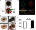
          Triple negative breast cancer (TNBC) is a belligerent carcinoma that is unresponsive to targeted receptor therapies. Development of new treatment strategies would benefit from an expanded repertoire of in vitro cell culture systems, such as those that support tridimensional growth in the presence of hydrogel scaffolds. To this end, we established protocols for maintenance of the TNBC cell line HCC70 in monolayer culture and in a commercially available basement membrane matrix hydrogel. We evaluated the general morphology of cells grown in both conditions with light microscopy, and examined their subcellular organization using transmission electron microscopy (TEM). Phase contrast and confocal microscopy showed the prevalence of irregularly shaped flattened cells in monolayer cultures, while cells maintained in hydrogel organized into multi-layered spheroids. A quantitative ultrastructural analysis comparing cells from the two culture conditions revealed that cells that formed spheroids comprised a greater number of mitochondria, autophagic vacuoles and intercellular junctions than their monolayer counterparts, within the equivalent area of sampled tissue. These observations suggest that triple negative breast cancer cells in culture can alter their organelle content, as well as their morphology, in response to their microenvironment. Methods presented here may be useful for those who intend to image cell cultures with TEM, and for investigators who seek to implement diverse in vitro models in the search for therapeutic molecular targets for TNBC.

          Related collections

          Most cited references47

          • Record: found
          • Abstract: found
          • Article: not found

          NIH Image to ImageJ: 25 years of image analysis.

          For the past 25 years NIH Image and ImageJ software have been pioneers as open tools for the analysis of scientific images. We discuss the origins, challenges and solutions of these two programs, and how their history can serve to advise and inform other software projects.
            Bookmark
            • Record: found
            • Abstract: found
            • Article: found
            Is Open Access

            3D tumor spheroid models for in vitro therapeutic screening: a systematic approach to enhance the biological relevance of data obtained

            The potential of a spheroid tumor model composed of cells in different proliferative and metabolic states for the development of new anticancer strategies has been amply demonstrated. However, there is little or no information in the literature on the problems of reproducibility of data originating from experiments using 3D models. Our analyses, carried out using a novel open source software capable of performing an automatic image analysis of 3D tumor colonies, showed that a number of morphology parameters affect the response of large spheroids to treatment. In particular, we found that both spheroid volume and shape may be a source of variability. We also compared some commercially available viability assays specifically designed for 3D models. In conclusion, our data indicate the need for a pre-selection of tumor spheroids of homogeneous volume and shape to reduce data variability to a minimum before use in a cytotoxicity test. In addition, we identified and validated a cytotoxicity test capable of providing meaningful data on the damage induced in large tumor spheroids of up to diameter in 650 μm by different kinds of treatments.
              Bookmark
              • Record: found
              • Abstract: found
              • Article: not found

              Tumor microenvironment: the role of the tumor stroma in cancer.

              The tumor microenvironment, composed of non-cancer cells and their stroma, has become recognized as a major factor influencing the growth of cancer. The microenvironment has been implicated in the regulation of cell growth, determining metastatic potential and possibly determining location of metastatic disease, and impacting the outcome of therapy. While the stromal cells are not malignant per se, their role in supporting cancer growth is so vital to the survival of the tumor that they have become an attractive target for chemotherapeutic agents. In this review, we will discuss the various cellular and molecular components of the stromal environment, their effects on cancer cell dynamics, and the rationale and implications of targeting this environment for control of cancer. Additionally, we will emphasize the role of the bone marrow-derived cell in providing cells for the stroma.
                Bookmark

                Author and article information

                Contributors
                Journal
                PeerJ
                PeerJ
                peerj
                peerj
                PeerJ
                PeerJ Inc. (San Francisco, USA )
                2167-8359
                16 February 2018
                2018
                : 6
                : e4340
                Affiliations
                [1 ]Department of Biology, New Mexico State University , Las Cruces, NM, United States of America
                Article
                4340
                10.7717/peerj.4340
                5817938
                29473000
                7259e96c-7159-4503-8d35-43933f40c763
                ©2018 Jogalekar and Serrano

                This is an open access article distributed under the terms of the Creative Commons Attribution License, which permits unrestricted use, distribution, reproduction and adaptation in any medium and for any purpose provided that it is properly attributed. For attribution, the original author(s), title, publication source (PeerJ) and either DOI or URL of the article must be cited.

                History
                : 28 June 2017
                : 18 January 2018
                Funding
                Funded by: NIH
                Award ID: P50GM068762
                Funded by: NSF
                Award ID: DBI-0959817
                Funded by: NMSU Manasse Endowment
                This research was supported by awards from NIH (P50GM068762), NSF (DBI-0959817) and the NMSU Manasse Endowment to EE Serrano. The funders had no role in study design, data collection and analysis, decision to publish, or preparation of the manuscript.
                Categories
                Cell Biology
                Anatomy and Physiology
                Oncology
                Pathology
                Histology

                autophagy,confocal microscopy,hydrogel,triple negative breast cancer,tissue engineering,phase contrast microscopy,ultrastructure,monolayer culture,tridimensional culture,transmission electron microscopy

                Comments

                Comment on this article