4
views
0
recommends
+1 Recommend
0 collections
    0
    shares
      • Record: found
      • Abstract: found
      • Article: found
      Is Open Access

      PKD1-associated autosomal dominant polycystic kidney disease with glomerular cysts presenting with nephrotic syndrome caused by focal segmental glomerulosclerosis

      case-report

      Read this article at

      Bookmark
          There is no author summary for this article yet. Authors can add summaries to their articles on ScienceOpen to make them more accessible to a non-specialist audience.

          Abstract

          Background

          Autosomal dominant polycystic kidney disease (ADPKD) may manifest non-nephrotic range proteinuria, but is rarely complicated with nephrotic syndrome. Limited number of reports describe the histology of ADPKD with nephrotic syndrome in detail.

          Case presentation

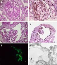
          We encountered a 23-year-old man with polycystic kidney disease (PKD) with small kidney volume and nephrotic syndrome, which eventually progressed to end-stage renal disease. Renal histology showed typical focal segmental glomerulosclerosis and remarkable glomerular cyst formation, but did not reveal tubular cysts. PKD1 mutation was detected in him and his father, who also had PKD with small kidney volume.

          Conclusions

          In contrast to tubular cysts which develop along ADPKD progression, glomerular cysts may likely be associated with ADPKD with slower volume progression manifesting small kidney volume. Although previous investigations report that ADPKD with smaller kidney volume is attributed to slower decline in renal function, coexistence of nephrotic-range proteinuria implies complication of other glomerular diseases and needs histological evaluation since it may lead to poor renal outcome.

          Related collections

          Most cited references8

          • Record: found
          • Abstract: found
          • Article: not found

          Imaging classification of autosomal dominant polycystic kidney disease: a simple model for selecting patients for clinical trials.

          The rate of renal disease progression varies widely among patients with autosomal dominant polycystic kidney disease (ADPKD), necessitating optimal patient selection for enrollment into clinical trials. Patients from the Mayo Clinic Translational PKD Center with ADPKD (n=590) with computed tomography/magnetic resonance images and three or more eGFR measurements over ≥6 months were classified radiologically as typical (n=538) or atypical (n=52). Total kidney volume (TKV) was measured using stereology (TKVs) and ellipsoid equation (TKVe). Typical patients were randomly partitioned into development and internal validation sets and subclassified according to height-adjusted TKV (HtTKV) ranges for age (1A-1E, in increasing order). Consortium for Radiologic Imaging Study of PKD (CRISP) participants (n=173) were used for external validation. TKVe correlated strongly with TKVs, without systematic underestimation or overestimation. A longitudinal mixed regression model to predict eGFR decline showed that log2HtTKV and age significantly interacted with time in typical patients, but not in atypical patients. When 1A-1E classifications were used instead of log2HtTKV, eGFR slopes were significantly different among subclasses and, except for 1A, different from those in healthy kidney donors. The equation derived from the development set predicted eGFR in both validation sets. The frequency of ESRD at 10 years increased from subclass 1A (2.4%) to 1E (66.9%) in the Mayo cohort and from 1C (2.2%) to 1E (22.3%) in the younger CRISP cohort. Class and subclass designations were stable. An easily applied classification of ADPKD based on HtTKV and age should optimize patient selection for enrollment into clinical trials and for treatment when one becomes available.
            Bookmark
            • Record: found
            • Abstract: found
            • Article: not found

            From segmental glomerulosclerosis to total nephron degeneration and interstitial fibrosis: a histopathological study in rat models and human glomerulopathies.

            Focal segmental glomerulosclerosis (FSGS) is consistently associated with tubular degeneration and interstitial fibrosis, altogether, accounting for the progressive decline in renal function. The mechanisms which link glomerular injury to tubulo-interstitial fibrosis are controversial. The present study describes the step-by-step sequence of histopathological events, i.e. the evolution of the injury from the initial lesion in the glomerulus to total nephron destruction. The investigation was performed in male hypertensive Fawn-hooded rats (6-, 9-, and 12-month-old) and 14-month-old Milan normotensive rats. The kidneys were fixed by in vivo perfusion and processed for structural investigation. Autopsy materials from human cases of focal segmental glomerulosclerosis and diabetic nephropathy were also examined. FSGS as seen in rat models consists of collapsed and hyalinized capillaries and mesangial portions which are included within a synechia between the glomerular tuft and Bowman's capsule. In addition, a synechia generally contains glomerular capillaries which are perfused and continue to filter with the filtrate being delivered into the interstitium rather than into Bowman's capsular space. Such filtration creates a paraglomerular space on the outer aspect of the parietal epithelium. This space becomes separated from the interstitium by a dense layer of sheet-like fibroblast processes. Associated with the progression to global sclerosis, this space spreads around the entire circumference of a glomerulus; all 'sclerotic' tuft portions are eventually contained in this space. Starting from the urinary pole this process also involves the proximal tubule, initially by expanding the tubular basement membrane (TBM) and later, by separating the TBM from its epithelium, thus creating a peritubular space by misdirected filtrate spreading. Similar to the situation observed at the glomerulus this space becomes separated from the interstitium by a layer of fibroblast processes. The final degeneration of the nephron occurs via two pathways. Pathway I whereby development to global sclerosis is dominant or develops concurrently with tubular degeneration, eventually terminating in global and cylindrical remnants of extracellular matrix surrounded by abundant fibrous tissue. Pathway II where the degeneration of the tubule is ahead of damage progression in the glomerulus leading to atubular glomerular cysts. The present study suggests that severely injured glomeruli may continue to filter with the filtrate spreading along interstitial routes. Fluid added locally to the interstitium from such 'extraterritorial' glomerular capillaries probably is quite different in quantity and composition compared to that from interstitial capillaries. We propose that this kind of abnormal addition of fluid to the interstitium is the essential mechanism accounting for interstitial progression of the disease. Similar histopathological phenomena in human kidneys with focal segmental glomerulosclerosis suggest that the pathogenetic pathways defined in the rat models operate in human disease as well.
              Bookmark
              • Record: found
              • Abstract: found
              • Article: not found

              Dietary protein restriction, blood pressure control, and the progression of polycystic kidney disease. Modification of Diet in Renal Disease Study Group.

              In the Modification of Diet in Renal Disease Study, a follow-up (mean, 2.2 yr) of 200 study participants with autosomal dominant polycystic kidney disease (ADPKD) was conducted to determine the effect of lowering protein intake and blood pressure on the rate of decline in GFR. The rate of decline was faster in participants with ADPKD than in persons with other diagnoses, reflecting, in part, faster disease progression in the ADPKD group. Baseline characteristics that predicted a faster rate of decline in GFR in persons with ADPKD were greater serum creatinine (independent of GFR), greater urinary protein excretion, higher mean arterial pressure (MAP), and younger age. In patients with initial GFR values between 25 and 55 mL/min per 1.73 m2, neither assignment to a low-protein diet group nor assignment to a low blood pressure group significantly reduced the rate of decline of GFR in ADPKD participants. Similarly, the decline in GFR was not related to achieved protein intake or MAP. In participants with GFR values between 13 and 24 mL/min per 1.73 m2, assignment to the low MAP group led to a somewhat more rapid decline in GFR. However, the more rapid decline in GFR did not appear to be due to a detrimental effect of low blood pressure or the antihypertensive agents used to reach the low blood pressure goal. Lower protein intake, but not prescription of the keto acid-amino acid supplement, was marginally associated with a slower progression of renal disease.
                Bookmark

                Author and article information

                Contributors
                +81-(0)3-3588-1111 , yasuhirooda3@gmail.com
                naokis@toranomon.gr.jp
                eiko-hase@hotmail.co.jp
                hilomiz@yahoo.co.jp
                kawada@toranomon.gr.jp
                akinari-s@toranomon.gr.jp
                rikako64@toranomon.gr.jp
                m.yamanouchi@toranomon.gr.jp
                non_love_99@yahoo.co.jp
                suwabetat@gmail.com
                hoshino@toranomon.gr.jp
                takaichi-tky@umin.ac.jp
                k.kinowaki@gmail.com
                ohashi@yokohama-cu.ac.jp
                tkshfj@gmail.com
                +81-(0)3-3588-1111 , ubara@toranomon.gr.jp
                Journal
                BMC Nephrol
                BMC Nephrol
                BMC Nephrology
                BioMed Central (London )
                1471-2369
                28 August 2019
                28 August 2019
                2019
                : 20
                : 337
                Affiliations
                [1 ]ISNI 0000 0004 1764 6940, GRID grid.410813.f, Nephrology Center, Toranomon Hospital, ; 2-2-2 Toranomon, Minato-ku, Tokyo, 105-8470 Japan
                [2 ]ISNI 0000 0004 1764 6940, GRID grid.410813.f, Okinaka Memorial Institute for Medical Research, Toranomon Hospital, ; 2-2-2 Toranomon, Minato-ku, Tokyo, 105-8470 Japan
                [3 ]ISNI 0000 0004 1764 6940, GRID grid.410813.f, Department of Pathology, , Toranomon Hospital, ; 2-2-2 Toranomon, Minato-ku, Tokyo, 105-8470 Japan
                [4 ]ISNI 0000 0001 1033 6139, GRID grid.268441.d, Department of Pathology, Graduate School of Medicine, , Yokohama City University, ; 3-9 Fukuura, Kanazawa-ku, Yokohama, Kanagawa 236-0004 Japan
                Author information
                http://orcid.org/0000-0002-7043-7676
                Article
                1524
                10.1186/s12882-019-1524-6
                6712641
                730d1bef-f1ed-48b3-a517-4f55003a25a5
                © The Author(s). 2019

                Open AccessThis article is distributed under the terms of the Creative Commons Attribution 4.0 International License ( http://creativecommons.org/licenses/by/4.0/), which permits unrestricted use, distribution, and reproduction in any medium, provided you give appropriate credit to the original author(s) and the source, provide a link to the Creative Commons license, and indicate if changes were made. The Creative Commons Public Domain Dedication waiver ( http://creativecommons.org/publicdomain/zero/1.0/) applies to the data made available in this article, unless otherwise stated.

                History
                : 20 May 2019
                : 16 August 2019
                Categories
                Case Report
                Custom metadata
                © The Author(s) 2019

                Nephrology
                autosomal dominant polycystic kidney disease,pkd1,glomerular cyst,focal segmental glomerulosclerosis

                Comments

                Comment on this article