10
views
0
recommends
+1 Recommend
0 collections
    0
    shares
      • Record: found
      • Abstract: found
      • Article: found
      Is Open Access

      Dental cell type atlas reveals stem and differentiated cell types in mouse and human teeth

      research-article

      Read this article at

      Bookmark
          There is no author summary for this article yet. Authors can add summaries to their articles on ScienceOpen to make them more accessible to a non-specialist audience.

          Abstract

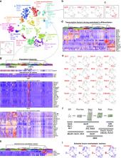
          Understanding cell types and mechanisms of dental growth is essential for reconstruction and engineering of teeth. Therefore, we investigated cellular composition of growing and non-growing mouse and human teeth. As a result, we report an unappreciated cellular complexity of the continuously-growing mouse incisor, which suggests a coherent model of cell dynamics enabling unarrested growth. This model relies on spatially-restricted stem, progenitor and differentiated populations in the epithelial and mesenchymal compartments underlying the coordinated expansion of two major branches of pulpal cells and diverse epithelial subtypes. Further comparisons of human and mouse teeth yield both parallelisms and differences in tissue heterogeneity and highlight the specifics behind growing and non-growing modes. Despite being similar at a coarse level, mouse and human teeth reveal molecular differences and species-specific cell subtypes suggesting possible evolutionary divergence. Overall, here we provide an atlas of human and mouse teeth with a focus on growth and differentiation.

          Abstract

          Unlike human teeth, mouse incisors grow throughout life, based on stem and progenitor cell activity. Here the authors generate single cell RNA-seq comparative maps of continuously-growing mouse incisor, non-growing mouse molar and human teeth, combined with lineage tracing to reveal dental cell complexity.

          Related collections

          Most cited references38

          • Record: found
          • Abstract: found
          • Article: not found

          Glial origin of mesenchymal stem cells in a tooth model system.

          Mesenchymal stem cells occupy niches in stromal tissues where they provide sources of cells for specialized mesenchymal derivatives during growth and repair. The origins of mesenchymal stem cells have been the subject of considerable discussion, and current consensus holds that perivascular cells form mesenchymal stem cells in most tissues. The continuously growing mouse incisor tooth offers an excellent model to address the origin of mesenchymal stem cells. These stem cells dwell in a niche at the tooth apex where they produce a variety of differentiated derivatives. Cells constituting the tooth are mostly derived from two embryonic sources: neural crest ectomesenchyme and ectodermal epithelium. It has been thought for decades that the dental mesenchymal stem cells giving rise to pulp cells and odontoblasts derive from neural crest cells after their migration in the early head and formation of ectomesenchymal tissue. Here we show that a significant population of mesenchymal stem cells during development, self-renewal and repair of a tooth are derived from peripheral nerve-associated glia. Glial cells generate multipotent mesenchymal stem cells that produce pulp cells and odontoblasts. By combining a clonal colour-coding technique with tracing of peripheral glia, we provide new insights into the dynamics of tooth organogenesis and growth.
            Bookmark
            • Record: found
            • Abstract: found
            • Article: not found

            Multilineage gene expression precedes commitment in the hemopoietic system.

            We have tested the hypothesis that multipotential hemopoietic stem and progenitor cells prime several different lineage-affiliated programs of gene activity prior to unilineage commitment and differentiation. Using single cell RT-PCR we show that erythroid (beta-globin) and myeloid (myeloperoxidase) gene expression programs can be initiated by the same cell prior to exclusive commitment to the erythroid or granulocytic lineages. Furthermore, the multipotential state is characterized by the coexpression of several lineage-affiliated cytokine receptors. These data support a model of hemopoietic lineage specification in which unilineage commitment is prefaced by a "promiscuous" phase of multilineage locus activation.
              Bookmark
              • Record: found
              • Abstract: found
              • Article: not found

              Dental mesenchymal stem cells.

              Mammalian teeth harbour mesenchymal stem cells (MSCs), which contribute to tooth growth and repair. These dental MSCs possess many in vitro features of bone marrow-derived MSCs, including clonogenicity, expression of certain markers, and following stimulation, differentiation into cells that have the characteristics of osteoblasts, chondrocytes and adipocytes. Teeth and their support tissues provide not only an easily accessible source of MSCs but also a tractable model system to study their function and properties in vivo In addition, the accessibility of teeth together with their clinical relevance provides a valuable opportunity to test stem cell-based treatments for dental disorders. This Review outlines some recent discoveries in dental MSC function and behaviour and discusses how these and other advances are paving the way for the development of new biologically based dental therapies.
                Bookmark

                Author and article information

                Contributors
                peter.kharchenko@post.harvard.edu
                igor.adameyko@ki.se
                Journal
                Nat Commun
                Nat Commun
                Nature Communications
                Nature Publishing Group UK (London )
                2041-1723
                23 September 2020
                23 September 2020
                2020
                : 11
                : 4816
                Affiliations
                [1 ]GRID grid.22937.3d, ISNI 0000 0000 9259 8492, Department of Molecular Neuroscience, Center for Brain Research, , Medical University of Vienna, ; Vienna, Austria
                [2 ]GRID grid.10267.32, ISNI 0000 0001 2194 0956, Department of Histology and Embryology, Faculty of Medicine, , Masaryk University, ; Brno, Czech Republic
                [3 ]GRID grid.38142.3c, ISNI 000000041936754X, Department of Biomedical Informatics, , Harvard Medical School, ; Boston, MA USA
                [4 ]GRID grid.4714.6, ISNI 0000 0004 1937 0626, Department of Physiology and Pharmacology, , Karolinska Institutet, ; Stockholm, Sweden
                [5 ]GRID grid.435109.a, ISNI 0000 0004 0639 4223, Institute of Animal Physiology and Genetics, CAS, ; Brno, Czech Republic
                [6 ]GRID grid.10267.32, ISNI 0000 0001 2194 0956, Clinic of Stomatology, Institution Shared with St. Anne’s Faculty Hospital, Faculty of Medicine, , Masaryk University, ; Brno, Czech Republic
                [7 ]GRID grid.22937.3d, ISNI 0000 0000 9259 8492, Department of Oral Biology, , Medical University of Vienna, ; Vienna, Austria
                [8 ]GRID grid.22937.3d, ISNI 0000 0000 9259 8492, Department of Oral Surgery, , Medical University of Vienna, ; Vienna, Austria
                [9 ]GRID grid.208078.5, ISNI 0000000419370394, Department of Craniofacial Sciences, School of Dental Medicine, , University of Connecticut Health Center, ; Farmington, CT USA
                [10 ]GRID grid.7737.4, ISNI 0000 0004 0410 2071, Research Program in Developmental Biology, Institute of Biotechnology, , University of Helsinki, ; Helsinki, Finland
                [11 ]GRID grid.266102.1, ISNI 0000 0001 2297 6811, Program in Craniofacial Biology and Department of Orofacial Sciences, , University of California, ; San Francisco, CA USA
                [12 ]GRID grid.266102.1, ISNI 0000 0001 2297 6811, Department of Pediatrics and Institute for Human Genetics, , University of California, ; San Francisco, CA USA
                [13 ]GRID grid.13097.3c, ISNI 0000 0001 2322 6764, Centre for Craniofacial and Regenerative Biology, Faculty of Dentistry, Oral & Craniofacial Sciences. King’s College London, ; London, UK
                [14 ]GRID grid.4714.6, ISNI 0000 0004 1937 0626, Department of Neuroscience, , Karolinska Institutet, ; Stockholm, Sweden
                [15 ]GRID grid.10420.37, ISNI 0000 0001 2286 1424, Department of Evolutionary Biology, , University of Vienna, ; Vienna, Austria
                [16 ]GRID grid.417850.f, ISNI 0000 0004 0639 5277, Centre d’Immunologie de Marseille-Luminy, Aix Marseille Université, INSERM, CNRS UMR, ; Marseille, France
                [17 ]GRID grid.22937.3d, ISNI 0000 0000 9259 8492, Department of Neuroimmunology, Center for Brain Research, , Medical University of Vienna, ; Vienna, Austria
                Author information
                http://orcid.org/0000-0002-7590-187X
                http://orcid.org/0000-0002-7444-0610
                http://orcid.org/0000-0002-7610-8929
                http://orcid.org/0000-0002-9311-8715
                http://orcid.org/0000-0001-6796-1197
                http://orcid.org/0000-0002-6254-7082
                http://orcid.org/0000-0001-9857-7577
                http://orcid.org/0000-0003-2116-9561
                http://orcid.org/0000-0002-6514-4406
                http://orcid.org/0000-0002-6036-5875
                http://orcid.org/0000-0001-5471-0356
                Article
                18512
                10.1038/s41467-020-18512-7
                7511944
                32968047
                74127a80-1590-447b-bf1c-bf29fc57b7e1
                © The Author(s) 2020

                Open Access This article is licensed under a Creative Commons Attribution 4.0 International License, which permits use, sharing, adaptation, distribution and reproduction in any medium or format, as long as you give appropriate credit to the original author(s) and the source, provide a link to the Creative Commons license, and indicate if changes were made. The images or other third party material in this article are included in the article’s Creative Commons license, unless indicated otherwise in a credit line to the material. If material is not included in the article’s Creative Commons license and your intended use is not permitted by statutory regulation or exceeds the permitted use, you will need to obtain permission directly from the copyright holder. To view a copy of this license, visit http://creativecommons.org/licenses/by/4.0/.

                History
                : 9 May 2020
                : 24 August 2020
                Funding
                Funded by: FundRef https://doi.org/10.13039/100011199, EC | EC Seventh Framework Programm | FP7 Ideas: European Research Council (FP7-IDEAS-ERC - Specific Programme: "Ideas" Implementing the Seventh Framework Programme of the European Community for Research, Technological Development and Demonstration Activities (2007 to 2013));
                Award ID: 647844
                Award Recipient :
                Categories
                Article
                Custom metadata
                © The Author(s) 2020

                Uncategorized
                organogenesis,stem-cell niche,mesenchymal stem cells
                Uncategorized
                organogenesis, stem-cell niche, mesenchymal stem cells

                Comments

                Comment on this article