24
views
0
recommends
+1 Recommend
0 collections
    0
    shares
      • Record: found
      • Abstract: found
      • Article: found
      Is Open Access

      Preliminary experience with cardiovascular magnetic resonance in evaluation of fetal cardiovascular anomalies

      research-article

      Read this article at

      Bookmark
          There is no author summary for this article yet. Authors can add summaries to their articles on ScienceOpen to make them more accessible to a non-specialist audience.

          Abstract

          Background

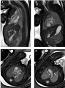
          The cardiovascular system is the part of the fetal anatomy that most frequently suffers from congenital pathology. This study shows our preliminary experience with fetal cardiovascular magnetic resonance (CMR) to evaluate congenital cardiovascular abnormalities.

          Methods

          Between January 2006 and June 2011, Prenatal routine obstetric ultrasound (US), echocardiography and CMR data from 68 pregnant women carrying fetuses with congenital cardiovascular anomalies were compared with postnatal diagnoses (postnatal imagings, surgery and autopsy). All prenatal CMR was performed at 1.5 T. Imaging sequences included steady-state free-precession (SSFP) sequences, real-time SSFP and single-shot turbo spin echo (SSTSE) sequences. The images were analyzed with an anatomic segmental approach by two radiologists.

          Results

          Fetal CMR yielded the same diagnosis as postnatal findings in 79% (54/68) of patients. The diagnostic sensitivity of routine obstetric US for cardiac anomalies was 46% (31/68). The diagnostic sensitivity of fetal echocardiographic examination by a fetal cardiac specialist was 82% (56/68). In 2 (3%) of 68 cases, diagnoses with both echocardiography and CMR were incorrect when compared with postnatal diagnosis. In ten (15%) cases, diagnosis at echocardiography was incorrect and that at CMR was correct. In twelve (18%) cases, diagnosis at echocardiography was correct and that at CMR was incorrect. Ten cases missed or misdiagnosed by echocardiography but correctly diagnosed by fetal CMR included asplenia syndrome (n = 2), interrupted inferior vena cava of polysplenia syndrome (n = 1), tricuspid incompetence (n = 1), double outlet right ventricle (n = 2), double aortic arch (n = 1), right pulmonary artery hypoplasia (n = 1), right-sided aortic arch of tetralogy of Fallot (n = 1) and hypoplastic left heart syndrome of a twin fetus (n = 1).

          Conclusion

          Fetal CMR is a promising diagnostic tool for assessment of congenital cardiovascular abnormalities, especially in situations that limit echocardiography.

          Related collections

          Most cited references23

          • Record: found
          • Abstract: found
          • Article: not found

          A review of the current use of magnetic resonance imaging in pregnancy and safety implications for the fetus.

          This paper presents an overview of the application of and risks of exposure to Magnetic Resonance Imaging (MRI) in pregnancy. It reviews the risks to the fetus by considering the hazards in terms of the three main components of an MRI system. These are the static magnetic field, the time-varying magnetic gradient fields and the pulsed radio frequency fields. The hazards discussed are biological effects, miscarriage, heating effects and acoustic noise exposure. This paper also presents a survey of MRI sites within the United Kingdom to ascertain the extent of MRI usage in pregnancy. To validate the situation of MRI in pregnancy a survey was sent to 352 MR units throughout the United Kingdom. The questions were grouped to assess (a) maternal MRI diagnosis (b) fetal MRI and (c) work practices for pregnant MRI staff. The results showed that 91% of sites were imaging pregnant women in need of diagnosis in the second and third trimester. This paper highlights that MRI can add information for fetal central nervous system abnormalities identified by ultrasound screening, however within the UK direct fetal imaging was only performed in 8% of sites. This paper indicates the need for research to be undertaken for specific MRI clinical conditions. It also advises that risk assessment for pregnant staff working in MRI is performed, and that there is a clear need for further research into the effects of MRI in pregnancy as there is a need for clear authoritive advice.
            Bookmark
            • Record: found
            • Abstract: found
            • Article: not found

            Prenatal ultrasound and fetal MRI: the comparative value of each modality in prenatal diagnosis.

            Fetal MRI is used with increasing frequency as an adjunct to ultrasound (US) in prenatal diagnosis. In this review, we discuss the relative value of both prenatal US and MRI in evaluating fetal and extra-fetal structures for a variety of clinical indications. Advantages and disadvantages of each imaging modality are addressed. In summary, MRI has advantages in demonstrating pathology of the brain, lungs, complex syndromes, and conditions associated with reduction of amniotic fluid. At present, US is the imaging method of choice during the first trimester, and in the diagnosis of cardiovascular abnormalities, as well as for screening. In some conditions, such as late gestational age, increased maternal body mass index, skeletal dysplasia, and metabolic disease, neither imaging method may provide sufficient diagnostic information.
              Bookmark
              • Record: found
              • Abstract: found
              • Article: not found

              MRI of normal and pathological fetal lung development.

              Normal fetal lung development is a complex process influenced by mechanical and many biochemical factors. In addition to ultrasound, fetal magnetic resonance imaging (MRI) constitutes a new method to investigate this process in vivo during the second and third trimester. The techniques of MRI volumetry, assessment of signal intensities, and MRI spectroscopy of the fetal lung have been used to analyze this process and have already been applied clinically to identify abnormal fetal lung growth. Particularly in conditions such as oligohydramnios and congenital diaphragmatic hernia (CDH), pulmonary hypoplasia may be the cause of neonatal death. A precise diagnosis and quantification of compromised fetal lung development may improve post- and perinatal management. The main events in fetal lung development are reviewed and MR volumetric data from 106 normal fetuses, as well as different examples of pathological lung growth, are provided.
                Bookmark

                Author and article information

                Contributors
                Journal
                J Cardiovasc Magn Reson
                J Cardiovasc Magn Reson
                Journal of Cardiovascular Magnetic Resonance
                BioMed Central
                1097-6647
                1532-429X
                2013
                21 May 2013
                : 15
                : 1
                : 40
                Affiliations
                [1 ]Department of Radiology, Shanghai Children’s Medical Center, Shanghai Jiaotong University School of Medicine, Shanghai 200127, China
                [2 ]Department of Cardiology, Shanghai Children’s Medical Center, Shanghai Jiaotong University School of Medicine, Shanghai 200127, China
                Article
                1532-429X-15-40
                10.1186/1532-429X-15-40
                3666966
                23692653
                742c3af0-07b6-45c1-bf34-ed1052af7bbb
                Copyright © 2013 Dong et al.; licensee BioMed Central Ltd.

                This is an Open Access article distributed under the terms of the Creative Commons Attribution License ( http://creativecommons.org/licenses/by/2.0), which permits unrestricted use, distribution, and reproduction in any medium, provided the original work is properly cited.

                History
                : 3 February 2012
                : 15 April 2013
                Categories
                Research

                Cardiovascular Medicine
                fetus,cardiovascular magnetic resonance,congenital heart disease,cardiac malformations,prenatal diagnosis

                Comments

                Comment on this article