0
views
0
recommends
+1 Recommend
0 collections
    0
    shares
      • Record: found
      • Abstract: found
      • Article: found
      Is Open Access

      3D-Cell-Annotator: an open-source active surface tool for single-cell segmentation in 3D microscopy images

      brief-report

      Read this article at

      Bookmark
          There is no author summary for this article yet. Authors can add summaries to their articles on ScienceOpen to make them more accessible to a non-specialist audience.

          Abstract

          Summary

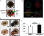
          Segmentation of single cells in microscopy images is one of the major challenges in computational biology. It is the first step of most bioimage analysis tasks, and essential to create training sets for more advanced deep learning approaches. Here, we propose 3D-Cell-Annotator to solve this task using 3D active surfaces together with shape descriptors as prior information in a semi-automated fashion. The software uses the convenient 3D interface of the widely used Medical Imaging Interaction Toolkit (MITK). Results on 3D biological structures (e.g. spheroids, organoids and embryos) show that the precision of the segmentation reaches the level of a human expert.

          Availability and implementation

          3D-Cell-Annotator is implemented in CUDA/C++ as a patch for the segmentation module of MITK. The 3D-Cell-Annotator enabled MITK distribution can be downloaded at: www.3D-cell-annotator.org. It works under Windows 64-bit systems and recent Linux distributions even on a consumer level laptop with a CUDA-enabled video card using recent NVIDIA drivers.

          Supplementary information

          Supplementary data are available at Bioinformatics online.

          Related collections

          Most cited references5

          • Record: found
          • Abstract: found
          • Article: found
          Is Open Access

          3D tumor spheroid models for in vitro therapeutic screening: a systematic approach to enhance the biological relevance of data obtained

          The potential of a spheroid tumor model composed of cells in different proliferative and metabolic states for the development of new anticancer strategies has been amply demonstrated. However, there is little or no information in the literature on the problems of reproducibility of data originating from experiments using 3D models. Our analyses, carried out using a novel open source software capable of performing an automatic image analysis of 3D tumor colonies, showed that a number of morphology parameters affect the response of large spheroids to treatment. In particular, we found that both spheroid volume and shape may be a source of variability. We also compared some commercially available viability assays specifically designed for 3D models. In conclusion, our data indicate the need for a pre-selection of tumor spheroids of homogeneous volume and shape to reduce data variability to a minimum before use in a cytotoxicity test. In addition, we identified and validated a cytotoxicity test capable of providing meaningful data on the damage induced in large tumor spheroids of up to diameter in 650 μm by different kinds of treatments.
            Bookmark
            • Record: found
            • Abstract: found
            • Article: not found

            The Medical Imaging Interaction Toolkit: challenges and advances : 10 years of open-source development.

            The Medical Imaging Interaction Toolkit (MITK) has been available as open-source software for almost 10 years now. In this period the requirements of software systems in the medical image processing domain have become increasingly complex. The aim of this paper is to show how MITK evolved into a software system that is able to cover all steps of a clinical workflow including data retrieval, image analysis, diagnosis, treatment planning, intervention support, and treatment control.
              Bookmark
              • Record: found
              • Abstract: found
              • Article: not found

              NucleusJ: an ImageJ plugin for quantifying 3D images of interphase nuclei.

              NucleusJ is a simple and user-friendly ImageJ plugin dedicated to the characterization of nuclear morphology and chromatin organization in 3D. Starting from image stacks, the nuclear boundary is delimited by combining the Otsu segmentation method with optimization of nuclear sphericity. Chromatin domains are segmented by partitioning the nucleus using a 3D watershed algorithm and by thresholding a contrast measure over the resulting regions. As output, NucleusJ quantifies 15 parameters including shape and size of nuclei as well as intra-nuclear objects and their position within the nucleus. A step-by-step documentation is available for self-training, together with data sets of nuclei with different nuclear organization.
                Bookmark

                Author and article information

                Contributors
                Role: Associate Editor
                Journal
                Bioinformatics
                Bioinformatics
                bioinformatics
                Bioinformatics
                Oxford University Press
                1367-4803
                1367-4811
                01 May 2020
                17 January 2020
                17 January 2020
                : 36
                : 9
                : 2948-2949
                Affiliations
                [b1 ] Synthetic and System Biology Unit , Biological Research Centre (BRC), Szeged H-6726, Hungary
                [b2 ] Buchmann Institute for Molecular Life Sciences (BMLS) , Goethe University of Frankfurt, DE-60438 Frankfurt, Germany
                [b3 ] Istituto Scientifico Romagnolo per lo Studio e la Cura dei Tumori (IRST) IRCCS , I-47014 Meldola (FC), Italy
                [b4 ] Institute for Molecular Medicine Finland University of Helsinki , FI-00014 Helsinki, Finland
                [b5 ] Single-Cell Technologies Ltd , H-6726 Szeged, Hungary
                Author notes
                To whom correspondence should be addressed. filippo.piccinini@ 123456irst.emr.it or horvath.peter@ 123456brc.hu
                Author information
                http://orcid.org/0000-0002-0371-7782
                Article
                btaa029
                10.1093/bioinformatics/btaa029
                7203751
                31950986
                78fd5bc7-8c20-4cc6-8aea-d0713dea71b2
                © The Author(s) 2020. Published by Oxford University Press.

                This is an Open Access article distributed under the terms of the Creative Commons Attribution Non-Commercial License ( http://creativecommons.org/licenses/by-nc/4.0/), which permits non-commercial re-use, distribution, and reproduction in any medium, provided the original work is properly cited. For commercial re-use, please contact journals.permissions@oup.com

                History
                : 23 June 2019
                : 30 December 2019
                : 15 January 2020
                Page count
                Pages: 2
                Funding
                Funded by: LENDULET-BIOMAG;
                Award ID: 2018-342
                Funded by: European Regional Development Funds;
                Award ID: GINOP-2.3.2-15-2016-00026
                Award ID: GINOP-2.3.2-15-2016-00037
                Funded by: Union for International Cancer Control, DOI 10.13039/100010260;
                Funded by: UICC, DOI 10.13039/100004432;
                Funded by: UICC Technical Fellowship;
                Award ID: UICC-TF/19/640197
                Categories
                Applications Notes
                Bioimage Informatics

                Bioinformatics & Computational biology
                Bioinformatics & Computational biology

                Comments

                Comment on this article