31
views
0
recommends
+1 Recommend
0 collections
    0
    shares
      • Record: found
      • Abstract: found
      • Article: found
      Is Open Access

      Clinical Presentation and Outcome of Sinonasal Tumors in a Nigerian Tertiary Hospital – 6-year Review

      research-article

      Read this article at

      Bookmark
          There is no author summary for this article yet. Authors can add summaries to their articles on ScienceOpen to make them more accessible to a non-specialist audience.

          Abstract

          Background:

          Sinonasal tumor is an uncommon tumor in Nigeria, but the burden of the disease in terms of morbidity and mortality is on the increase. The aim of this study is to evaluate the pattern of presentation and outcome of sinonasal tumor in our center.

          Methodology:

          This was a retrospective review of all the patients with sinonasal cancer seen in Ear, Nose, and Throat Department of the hospital between January 2008 and December 2013. The patients' biodata, clinical presentation, stage of the disease at presentation, identifiable risk factors, histopathological findings, and outcome were presented.

          Results:

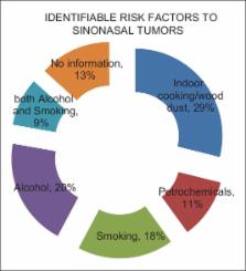
          A total of 22 patients were diagnosed with sinonasal cancer out of 71 patients (31%) with head/neck cancers during the period of study. There were 12 males (54.5%) and 10 females (45.5%) with a mean age of 51 ± 2.1 years. Epistaxis, nasal blockage, and discharge were seen in all patients though 41% of patients presented first to the ophthalmologist due to proptosis. Identifiable risk factors were exposure to wood dust (29%) and petrochemical products in (11%). Most patients presented first at advanced stages of the disease (82%). Histologically, well-differentiated squamous cell carcinoma was the most common (54.6%) followed by nonintestinal well-differentiated adenocarcinoma in 18.2% and plasmacytoma (9.1%). Chemoradiation was the mainstay of management in (38%) though 13.6% had total maxillectomy with postsurgical chemoradiation and 50.4% defaulted due to cost of management.

          Conclusion:

          Health education on early presentation and efforts at early detection of the disease are needed to achieve cure. Exposure to the identifiable risk factors should be reduced by protective measures. Easy access to radiotherapy at affordable price will surely improve the outcome of this disease.

          Related collections

          Most cited references19

          • Record: found
          • Abstract: found
          • Article: not found

          Craniofacial resection for malignant paranasal sinus tumors: Report of an International Collaborative Study.

          Malignant tumors of the superior sinonasal vault are rare, and, because of this and the varied histologic findings, most outcomes data reflect the experience of small patient cohorts. This International Collaborative study examines a large cohort of patients accumulated from multiple institutions experienced in craniofacial surgery, with the aim of reporting benchmark figures for outcomes and identifying patient-related and tumor-related predictors of prognosis after craniofacial resection (CFR). Three hundred thirty-four patients from 17 institutions were analyzed for outcome. Patients with esthesioneuroblastoma were excluded and are being reported separately. The median age was 57 years (range, 3-98 years). One hundred eighty-eight patients (56.3%) had had prior single-modality or combined treatment, which included surgery in 120 (36%), radiation in 79 (23.7%), and chemotherapy in 56 (16.8%). The most common histologic findings were adenocarcinoma in 107 (32%) and squamous cell carcinoma in 101 (30.2%). The margins of resection were close or microscopically positive in 95 (30%). Adjuvant radiotherapy was given in 161 (48.2%) and chemotherapy in 16 (4.8%). Statistical analyses for outcomes were performed in relation to patient characteristics, tumor characteristics, including histologic findings and extent of disease, surgical resection margins, prior radiation, and prior chemotherapy to determine predictive factors. Postoperative mortality occurred in 15 patients (4.5%). Postoperative complications occurred in 110 patients (32.9%). The 5-year overall, disease-specific, and recurrence-free survival rates were 48.3%, 53.3%, and 45.8%, respectively. The status of surgical margins, histologic findings of the primary tumor, and intracranial extent were independent predictors of overall, disease-specific, and recurrence-free survival on multivariate analysis. CFR for malignant paranasal sinus tumors is a safe surgical treatment with an overall mortality of 4.5% and complication rate of 33%. The status of surgical margins, histologic findings of the primary tumor, and intracranial extent are independent predictors of outcome.
            Bookmark
            • Record: found
            • Abstract: found
            • Article: not found

            Management of the orbit in malignant sinonasal tumors.

            Malignant ethmoid and maxillary sinus tumors frequently involve the orbit. Orbital involvement is an important prognostic predictor of recurrence-free, disease-specific, and overall survival. Most authors agree that orbital preservation as opposed to orbital exenteration or clearance does not result in significant differences in local recurrence or actuarial survival. The eye can be safely preserved in most patients with ethmoid or maxillary sinus cancer invading the orbital wall, including malignancies that invade the orbital soft tissues with penetration through the periorbita provided that they can be completely dissected away from the orbital fat. Malposition of the globe and nonfunctional eyes frequently result when patients have not had adequate rigid reconstruction of the orbital floor, particularly if they have received postoperative radiotherapy. This underscores the importance of such reconstruction. Isolated defects following orbital exenteration may be reconstructed with a temporalis muscle flap. Microvascular free-tissue transfer is the best option for repair of defects following orbital exenteration and total maxillectomy, although an obturator still has a role in selected patients.
              Bookmark
              • Record: found
              • Abstract: found
              • Article: not found

              Ethmoido-Orbital Tumors: Our Experience

              No data in the literature report the specific invasion of the orbit from ethmoidal tumors, although such a pattern of involvement of the orbit frequently occurs because of the thin lamina papyracea separating the two structures. From January 1986 to January 2003, 38 patients with untreated primary ethmoidal malignancies were observed at the Unit of Maxillo-Facial Surgery of the University "La Sapienza" in Rome. Personal data were obtained from review of the personal clinical file of each. Orbital invasion was present in 24 patients with ethmoidal malignancy. Three stages of orbital invasion were identified. The average overall survival, with the Kaplan-Meyer method, was 61.4% after 1 year and 51.1% at 5 and 10 years. Intracranial involvement is the main element for short-term negative prognosis. Orbital exenteration is fundamental if grade III orbital invasion occurs because it ensures an improvement of the disease-free survival.
                Bookmark

                Author and article information

                Journal
                Niger Med J
                Niger Med J
                NMJ
                Nigerian Medical Journal : Journal of the Nigeria Medical Association
                Medknow Publications & Media Pvt Ltd (India )
                0300-1652
                2229-774X
                May-Jun 2017
                : 58
                : 3
                : 92-95
                Affiliations
                [1]Department of Ear, Nose and Throat, University of Ilorin/University of Ilorin Teaching Hospital, Ilorin, Nigeria
                [1 ]Department of ORL, University of Ilorin/University of Ilorin Teaching Hospital, Ilorin, Nigeria
                Author notes
                Address for correspondence: Dr. B. S. Alabi, Department of ORL, University of Ilorin Teaching Hospital, University of Ilorin, Ilorin, Nigeria. E-mail: alabibs@ 123456yahoo.com
                Article
                NMJ-58-92
                10.4103/nmj.NMJ_265_14
                6009142
                29962649
                7a34ab88-936e-4bd2-974a-7d3eac82986c
                Copyright: © 2018 Nigerian Medical Journal

                This is an open access journal, and articles are distributed under the terms of the Creative Commons Attribution-NonCommercial-ShareAlike 4.0 License, which allows others to remix, tweak, and build upon the work non-commercially, as long as appropriate credit is given and the new creations are licensed under the identical terms.

                History
                Categories
                Original Article

                Medicine
                outcome,pathology,presentation,risk factors,sinonasal tumor
                Medicine
                outcome, pathology, presentation, risk factors, sinonasal tumor

                Comments

                Comment on this article