12
views
0
recommends
+1 Recommend
0 collections
    0
    shares
      • Record: found
      • Abstract: found
      • Article: found
      Is Open Access

      Roles of sarcoplasmic reticulum Ca 2+ ATPase pump in the impairments of lymphatic contractile activity in a metabolic syndrome rat model

      research-article

      Read this article at

      Bookmark
          There is no author summary for this article yet. Authors can add summaries to their articles on ScienceOpen to make them more accessible to a non-specialist audience.

          Abstract

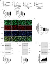
          The intrinsic lymphatic contractile activity is necessary for proper lymph transport. Mesenteric lymphatic vessels from high-fructose diet-induced metabolic syndrome (MetSyn) rats exhibited impairments in its intrinsic phasic contractile activity; however, the molecular mechanisms responsible for the weaker lymphatic pumping activity in MetSyn conditions are unknown. Several metabolic disease models have shown that dysregulation of sarcoplasmic reticulum Ca 2+ ATPase (SERCA) pump is one of the key determinants of the phenotypes seen in various muscle tissues. Hence, we hypothesized that a decrease in SERCA pump expression and/or activity in lymphatic muscle influences the diminished lymphatic vessel contractions in MetSyn animals. Results demonstrated that SERCA inhibitor, thapsigargin, significantly reduced lymphatic phasic contractile frequency and amplitude in control vessels, whereas, the reduced MetSyn lymphatic contractile activity was not further diminished by thapsigargin. While SERCA2a expression was significantly decreased in MetSyn lymphatic vessels, myosin light chain 20, MLC 20 phosphorylation was increased in these vessels. Additionally, insulin resistant lymphatic muscle cells exhibited elevated intracellular calcium and decreased SERCA2a expression and activity. The SERCA activator, CDN 1163 partially restored lymphatic contractile activity in MetSyn lymphatic vessel by increasing phasic contractile frequency. Thus, our data provide the first evidence that SERCA2a modulates the lymphatic pumping activity by regulating phasic contractile amplitude and frequency, but not the lymphatic tone. Diminished lymphatic contractile activity in the vessels from the MetSyn animal is associated with the decreased SERCA2a expression and impaired SERCA2 activity in lymphatic muscle.

          Related collections

          Most cited references73

          • Record: found
          • Abstract: found
          • Article: not found

          Ca2+ sensitivity of smooth muscle and nonmuscle myosin II: modulated by G proteins, kinases, and myosin phosphatase.

          Ca2+ sensitivity of smooth muscle and nonmuscle myosin II reflects the ratio of activities of myosin light-chain kinase (MLCK) to myosin light-chain phosphatase (MLCP) and is a major, regulated determinant of numerous cellular processes. We conclude that the majority of phenotypes attributed to the monomeric G protein RhoA and mediated by its effector, Rho-kinase (ROK), reflect Ca2+ sensitization: inhibition of myosin II dephosphorylation in the presence of basal (Ca2+ dependent or independent) or increased MLCK activity. We outline the pathway from receptors through trimeric G proteins (Galphaq, Galpha12, Galpha13) to activation, by guanine nucleotide exchange factors (GEFs), from GDP. RhoA. GDI to GTP. RhoA and hence to ROK through a mechanism involving association of GEF, RhoA, and ROK in multimolecular complexes at the lipid cell membrane. Specific domains of GEFs interact with trimeric G proteins, and some GEFs are activated by Tyr kinases whose inhibition can inhibit Rho signaling. Inhibition of MLCP, directly by ROK or by phosphorylation of the phosphatase inhibitor CPI-17, increases phosphorylation of the myosin II regulatory light chain and thus the activity of smooth muscle and nonmuscle actomyosin ATPase and motility. We summarize relevant effects of p21-activated kinase, LIM-kinase, and focal adhesion kinase. Mechanisms of Ca2+ desensitization are outlined with emphasis on the antagonism between cGMP-activated kinase and the RhoA/ROK pathway. We suggest that the RhoA/ROK pathway is constitutively active in a number of organs under physiological conditions; its aberrations play major roles in several disease states, particularly impacting on Ca2+ sensitization of smooth muscle in hypertension and possibly asthma and on cancer neoangiogenesis and cancer progression. It is a potentially important therapeutic target and a subject for translational research.
            Bookmark
            • Record: found
            • Abstract: found
            • Article: not found

            NADPH oxidase 4 (Nox4) is a major source of oxidative stress in the failing heart.

            NAD(P)H oxidases (Noxs) produce O(2)(-) and play an important role in cardiovascular pathophysiology. The Nox4 isoform is expressed primarily in the mitochondria in cardiac myocytes. To elucidate the function of endogenous Nox4 in the heart, we generated cardiac-specific Nox4(-/-) (c-Nox4(-/-)) mice. Nox4 expression was inhibited in c-Nox4(-/-) mice in a heart-specific manner, and there was no compensatory up-regulation in other Nox enzymes. These mice exhibited reduced levels of O(2)(-) in the heart, indicating that Nox4 is a significant source of O(2)(-) in cardiac myocytes. The baseline cardiac phenotype was normal in young c-Nox4(-/-) mice. In response to pressure overload (PO), however, increases in Nox4 expression and O(2)(-) production in mitochondria were abolished in c-Nox4(-/-) mice, and c-Nox4(-/-) mice exhibited significantly attenuated cardiac hypertrophy, interstitial fibrosis and apoptosis, and better cardiac function compared with WT mice. Mitochondrial swelling, cytochrome c release, and decreases in both mitochondrial DNA and aconitase activity in response to PO were attenuated in c-Nox4(-/-) mice. On the other hand, overexpression of Nox4 in mouse hearts exacerbated cardiac dysfunction, fibrosis, and apoptosis in response to PO. These results suggest that Nox4 in cardiac myocytes is a major source of mitochondrial oxidative stress, thereby mediating mitochondrial and cardiac dysfunction during PO.
              Bookmark
              • Record: found
              • Abstract: found
              • Article: not found

              Lymphatic vascular defects promoted by Prox1 haploinsufficiency cause adult-onset obesity.

              Multiple organs cooperate to regulate appetite, metabolism, and glucose and fatty acid homeostasis. Here, we identified and characterized lymphatic vasculature dysfunction as a cause of adult-onset obesity. We found that functional inactivation of a single allele of the homeobox gene Prox1 led to adult-onset obesity due to abnormal lymph leakage from mispatterned and ruptured lymphatic vessels. Prox1 heterozygous mice are a new model for adult-onset obesity and lymphatic vascular disease.
                Bookmark

                Author and article information

                Contributors
                marim@tamu.edu
                Journal
                Sci Rep
                Sci Rep
                Scientific Reports
                Nature Publishing Group UK (London )
                2045-2322
                23 July 2020
                23 July 2020
                2020
                : 10
                : 12320
                Affiliations
                [1 ]ISNI 0000 0004 4687 2082, GRID grid.264756.4, Department of Medical Physiology, College of Medicine, , Texas A&M University, ; Bryan, TX 77807 USA
                [2 ]Present Address: Vascular Biology Program, Boston Children’s Hospital, Harvard Medical School, Boston, MA 02115 USA
                Article
                69196
                10.1038/s41598-020-69196-4
                7378550
                32704072
                7a8ea6f1-a465-4570-ab5b-d75d637dfa45
                © The Author(s) 2020

                Open Access This article is licensed under a Creative Commons Attribution 4.0 International License, which permits use, sharing, adaptation, distribution and reproduction in any medium or format, as long as you give appropriate credit to the original author(s) and the source, provide a link to the Creative Commons license, and indicate if changes were made. The images or other third party material in this article are included in the article’s Creative Commons license, unless indicated otherwise in a credit line to the material. If material is not included in the article’s Creative Commons license and your intended use is not permitted by statutory regulation or exceeds the permitted use, you will need to obtain permission directly from the copyright holder. To view a copy of this license, visit http://creativecommons.org/licenses/by/4.0/.

                History
                : 10 January 2020
                : 9 July 2020
                Funding
                Funded by: NIH NIDDK RO1 DK99221
                Categories
                Article
                Custom metadata
                © The Author(s) 2020

                Uncategorized
                cell biology,molecular biology,physiology
                Uncategorized
                cell biology, molecular biology, physiology

                Comments

                Comment on this article