36
views
0
recommends
+1 Recommend
0 collections
    0
    shares
      • Record: found
      • Abstract: found
      • Article: found
      Is Open Access

      Recessive nephrocerebellar syndrome on the Galloway-Mowat syndrome spectrum is caused by homozygous protein-truncating mutations of WDR73

      research-article
      1 , , 1 , 2 , 3 , 4 , 5 , 6 , 7 , 8 , 9 , 10 , 11 , 1 , 1 , 1 , 1 , 1 , 12 , 13 , 14 , 13 , 12 , 13 , 2 , 2 , 15 , 16 , 17 , 18 , 18 , 19 , 20 , 1 , 2 , 21 , 11 , 22 , 3 , 1 , 2 , 21
      Brain
      Oxford University Press
      progressive microcephaly, nephrosis, cerebellar hypoplasia, mitosis, mTOR

      Read this article at

      Bookmark
          There is no author summary for this article yet. Authors can add summaries to their articles on ScienceOpen to make them more accessible to a non-specialist audience.

          Abstract

          Galloway-Mowat syndrome (GMS) is a neurodevelopmental disorder characterized by microcephaly, cerebellar hypoplasia, nephrosis, and profound intellectual disability. Jinks et al. extend the GMS spectrum by identifying a novel nephrocerebellar syndrome with selective striatal cholinergic interneuron loss and complete lateral geniculate nucleus delamination, caused by a frameshift mutation in WDR73.

          Abstract

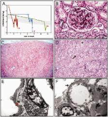
          We describe a novel nephrocerebellar syndrome on the Galloway-Mowat syndrome spectrum among 30 children (ages 1.0 to 28 years) from diverse Amish demes. Children with nephrocerebellar syndrome had progressive microcephaly, visual impairment, stagnant psychomotor development, abnormal extrapyramidal movements and nephrosis. Fourteen died between ages 2.7 and 28 years, typically from renal failure. Post-mortem studies revealed (i) micrencephaly without polymicrogyria or heterotopia; (ii) atrophic cerebellar hemispheres with stunted folia, profound granule cell depletion, Bergmann gliosis, and signs of Purkinje cell deafferentation; (iii) selective striatal cholinergic interneuron loss; and (iv) optic atrophy with delamination of the lateral geniculate nuclei. Renal tissue showed focal and segmental glomerulosclerosis and extensive effacement and microvillus transformation of podocyte foot processes. Nephrocerebellar syndrome mapped to 700 kb on chromosome 15, which contained a single novel homozygous frameshift variant ( WDR73 c.888delT; p.Phe296Leufs*26). WDR73 protein is expressed in human cerebral cortex, hippocampus, and cultured embryonic kidney cells. It is concentrated at mitotic microtubules and interacts with α-, β-, and γ-tubulin, heat shock proteins 70 and 90 (HSP-70; HSP-90), and the carbamoyl phosphate synthetase 2/aspartate transcarbamylase/dihydroorotase multi-enzyme complex. Recombinant WDR73 p.Phe296Leufs*26 and p.Arg256Profs*18 proteins are truncated, unstable, and show increased interaction with α- and β-tubulin and HSP-70/HSP-90. Fibroblasts from patients homozygous for WDR73 p.Phe296Leufs*26 proliferate poorly in primary culture and senesce early. Our data suggest that in humans, WDR73 interacts with mitotic microtubules to regulate cell cycle progression, proliferation and survival in brain and kidney. We extend the Galloway-Mowat syndrome spectrum with the first description of diencephalic and striatal neuropathology.

          Related collections

          Most cited references59

          • Record: found
          • Abstract: found
          • Article: not found

          Stimulation of de novo pyrimidine synthesis by growth signaling through mTOR and S6K1.

          Cellular growth signals stimulate anabolic processes. The mechanistic target of rapamycin complex 1 (mTORC1) is a protein kinase that senses growth signals to regulate anabolic growth and proliferation. Activation of mTORC1 led to the acute stimulation of metabolic flux through the de novo pyrimidine synthesis pathway. mTORC1 signaling posttranslationally regulated this metabolic pathway via its downstream target ribosomal protein S6 kinase 1 (S6K1), which directly phosphorylates S1859 on CAD (carbamoyl-phosphate synthetase 2, aspartate transcarbamoylase, dihydroorotase), the enzyme that catalyzes the first three steps of de novo pyrimidine synthesis. Growth signaling through mTORC1 thus stimulates the production of new nucleotides to accommodate an increase in RNA and DNA synthesis needed for ribosome biogenesis and anabolic growth.
            Bookmark
            • Record: found
            • Abstract: found
            • Article: not found

            Whole exome sequencing identifies recessive WDR62 mutations in severe brain malformations

            The development of the human cerebral cortex is an orchestrated process involving the birth of neural progenitors in the peri-ventricular germinal zones, cell proliferation characterized by both symmetric and asymmetric mitoses, followed by migration of post-mitotic neurons to their final destinations in 6 highly ordered, functionally-specialized layers 1,2 . An understanding of the molecular mechanisms guiding these intricate processes is in its infancy, substantially driven by the discovery of rare mutations that cause malformations of cortical development (MCD) 3-6 . Mapping of disease loci in putative Mendelian forms of MCD has been hindered by marked locus heterogeneity, small kindred sizes and diagnostic classifications that may not reflect molecular pathogenesis. Here we demonstrate the use of whole-exome sequencing to overcome these obstacles by identifying recessive mutations in WDR62 as the cause of a wide spectrum of severe cerebral cortical malformations including microcephaly, pachygria with cortical thickening as well as hypoplasia of the corpus callosum. Some patients with WDR62 mutations had evidence of additional abnormalities including lissencephaly, schizencephaly, polymicrogyria and, in one instance, cerebellar hypoplasia, all traits traditionally regarded as distinct entities. In mouse and humans, WDR62 transcripts and protein are enriched in neural progenitors within the ventricular and subventricular zones. WDR62 expression in the neocortex is transient, spanning the period of embryonic neurogenesis. Unlike other known microcephaly genes, WDR62 does not apparently associate with centrosomes and is predominantly nuclear in localization. These findings unify previously disparate aspects of cerebral cortical development and highlight the utility of whole-exome sequencing to identify disease loci in settings in which traditional methods have proved challenging.
              Bookmark
              • Record: found
              • Abstract: found
              • Article: not found

              Cerebellum of the premature infant: rapidly developing, vulnerable, clinically important.

              J Volpe (2009)
              Brain abnormality in surviving premature infants is associated with an enormous amount of neurodevelopmental disability, manifested principally by cognitive, behavioral, attentional, and socialization deficits, most commonly with only relatively modest motor deficits. The most recognized contributing neuropathology is cerebral white matter injury. The thesis of this review is that acquired cerebellar abnormality is a relatively less recognized but likely important cause of neurodevelopmental disability in small premature infants. The cerebellar disease may be primarily destructive (eg, hemorrhage, infarction) or primarily underdevelopment. The latter appears to be especially common and relates to a particular vulnerability of the cerebellum of the small premature infant. Central to this vulnerability are the extraordinarily rapid and complex developmental events occurring in the cerebellum. The disturbance of development can be caused either by direct adverse effects on the cerebellum, especially the distinctive transient external granular layer, or by indirect remote trans-synaptic effects. This review describes the fascinating details of cerebellar development, with an emphasis on events in the premature period, the major types of cerebellar abnormality acquired during the premature period, their likely mechanisms of occurrence, and new insights into the relation of cerebellar disease in early life to subsequent cognitive/behavioral/attentional/socialization deficits.
                Bookmark

                Author and article information

                Journal
                Brain
                Brain
                brainj
                brain
                Brain
                Oxford University Press
                0006-8950
                1460-2156
                August 2015
                12 June 2015
                12 June 2015
                : 138
                : 8
                : 2173-2190
                Affiliations
                1 Department of Biology and Biological Foundations of Behaviour Program, Franklin and Marshall College, Lancaster, PA 17604, USA
                2 Clinic for Special Children, Strasburg, PA 17579, USA
                3 RILD Wellcome Wolfson Centre, Royal Devon and Exeter NHS Foundation Trust, Barrack Road, Exeter, EX2 5DW, UK
                4 Human Genetics and Genomic Medicine, Faculty of Medicine, University of Southampton, UK
                5 Wessex Clinical Genetics Service, Princess Anne Hospital, Southampton, UK
                6 Department of Pathology and Laboratory Medicine, Children’s Hospital of Philadelphia and Perelman School of Medicine, University of Pennsylvania, Philadelphia, PA 19104, USA
                7 Shriners Hospital Paediatric Research Centre, Temple University School of Medicine, Philadelphia, PA 19140, USA
                8 Division of Renal Pathology, Vanderbilt University School of Medicine, Nashville, TN 37232, USA
                9 New Leaf Clinic for Special Children, Mount Eaton, OH 44659, USA
                10 Department of Paediatrics, Akron Children’s Hospital, Akron, OH 44302, USA
                11 DDC Clinic for Special Needs Children, Middlefield, OH 44062, USA
                12 Wellcome Trust Sanger Institute, Hinxton, Cambridge CB10 1SA, UK
                13 Medical Research, RILD Wellcome Wolfson Centre, University of Exeter Medical School, Exeter EX1 2LU, UK
                14 Department of Ophthalmology, University of Arizona College of Medicine, Tucson, AZ 85711, USA
                15 Department of Pathology, University Hospitals Case Medical Centre, Cleveland, OH 44106, USA
                16 Case Western Reserve University School of Medicine, Cleveland, OH 44106, USA
                17 Howard Hughes Medical Institute, Chevy Chase, MD 20815, USA
                18 Division of Nephrology, Department of Medicine, Boston Children’s Hospital, Harvard Medical School, Boston, MA 02115, USA
                19 Department of Paediatrics, Nemours/Alfred I. DuPont Hospital for Children, Wilmington, DE 19803, USA
                20 Department of Molecular and Cell Biology and Institute for Systems Genomics, University of Connecticut, Storrs, CT 06269, USA
                21 Lancaster General Hospital, Lancaster, PA 17602, USA
                22 Department of Paediatrics, Rainbow Babies and Children’s Hospital and Department of Molecular Cardiology, Cleveland Clinic, Cleveland, OH 44195, USA
                Author notes
                Correspondence to: Robert N. Jinks, Ph.D., Department of Biology, Franklin and Marshall College, PO Box 3003, Lancaster, PA 17604-3003, USA E-mail: rjinks@ 123456fandm.edu
                Article
                awv153
                10.1093/brain/awv153
                4511861
                26070982
                7ae2e3b7-09fe-4e6f-8ff9-5386bc411efc
                © The Author (2015). Published by Oxford University Press on behalf of the Guarantors of Brain. All rights reserved. For Permissions, please email: journals.permissions@oup.com

                This is an Open Access article distributed under the terms of the Creative Commons Attribution License ( http://creativecommons.org/licenses/by/4.0/), which permits unrestricted reuse, distribution, and reproduction in any medium, provided the original work is properly cited.

                History
                : 05 December 2014
                : 19 March 2015
                : 14 April 2015
                Page count
                Pages: 18
                Categories
                Original Articles
                1030

                Neurosciences
                progressive microcephaly,nephrosis,cerebellar hypoplasia,mitosis,mtor
                Neurosciences
                progressive microcephaly, nephrosis, cerebellar hypoplasia, mitosis, mtor

                Comments

                Comment on this article