15
views
0
recommends
+1 Recommend
0 collections
    0
    shares
      • Record: found
      • Abstract: found
      • Article: found
      Is Open Access

      Genetics of Childhood Steroid Sensitive Nephrotic Syndrome: An Update

      review-article

      Read this article at

      Bookmark
          There is no author summary for this article yet. Authors can add summaries to their articles on ScienceOpen to make them more accessible to a non-specialist audience.

          Abstract

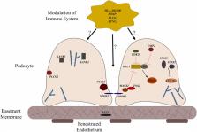
          Advances in genome science in the last 20 years have led to the discovery of over 50 single gene causes and genetic risk loci for steroid resistant nephrotic syndrome (SRNS). Despite these advances, the genetic architecture of childhood steroid sensitive nephrotic syndrome (SSNS) remains poorly understood due in large part to the varying clinical course of SSNS over time. Recent exome and genome wide association studies from well-defined cohorts of children with SSNS identified variants in multiple MHC class II molecules such as HLA-DQA1 and HLA-DQB1 as risk factors for SSNS, thus stressing the central role of adaptive immunity in the pathogenesis of SSNS. However, evidence suggests that unknown second hit risk loci outside of the MHC locus and environmental factors also make significant contributions to disease. In this review, we examine what is currently known about the genetics of SSNS, the implications of recent findings on our understanding of pathogenesis of SSNS, and how we can utilize these results and findings from future studies to improve the management of children with nephrotic syndrome.

          Related collections

          Most cited references97

          • Record: found
          • Abstract: found
          • Article: not found

          Idiopathic nephrotic syndrome in children

          The incidence of idiopathic nephrotic syndrome (NS) is 1·15-16·9 per 100 000 children, varying by ethnicity and region. The cause remains unknown but the pathogenesis of idiopathic NS is thought to involve immune dysregulation, systemic circulating factors, or inherited structural abnormalities of the podocyte. Genetic risk is more commonly described among children with steroid-resistant disease. The mainstay of therapy is prednisone for the vast majority of patients who are steroid responsive; however, the disease can run a frequently relapsing course, necessitating the need for alternative immunosuppressive agents. Infection and venous thromboembolism are the main complications of NS with also increased risk of acute kidney injury. Prognosis in terms of long-term kidney outcome overall is excellent for steroid-responsive disease, and steroid resistance is an important determinant of future risk of chronic or end-stage kidney disease.
            Bookmark
            • Record: found
            • Abstract: found
            • Article: not found

            Modification of kidney barrier function by the urokinase receptor.

            Podocyte dysfunction, represented by foot process effacement and proteinuria, is often the starting point for progressive kidney disease. Therapies aimed at the cellular level of the disease are currently not available. Here we show that induction of urokinase receptor (uPAR) signaling in podocytes leads to foot process effacement and urinary protein loss via a mechanism that includes lipid-dependent activation of alphavbeta3 integrin. Mice lacking uPAR (Plaur-/-) are protected from lipopolysaccharide (LPS)-mediated proteinuria but develop disease after expression of a constitutively active beta3 integrin. Gene transfer studies reveal a prerequisite for uPAR expression in podocytes, but not in endothelial cells, for the development of LPS-mediated proteinuria. Mechanistically, uPAR is required to activate alphavbeta3 integrin in podocytes, promoting cell motility and activation of the small GTPases Cdc42 and Rac1. Blockade of alphavbeta3 integrin reduces podocyte motility in vitro and lowers proteinuria in mice. Our findings show a physiological role for uPAR signaling in the regulation of kidney permeability.
              Bookmark
              • Record: found
              • Abstract: found
              • Article: not found

              Rituximab targets podocytes in recurrent focal segmental glomerulosclerosis.

              Focal segmental glomerulosclerosis (FSGS) is a glomerular disease characterized by proteinuria, progression to end-stage renal disease, and recurrence of proteinuria after kidney transplantation in about one-third of patients. It has been suggested that rituximab might treat recurrent FSGS through an unknown mechanism. Rituximab not only recognizes CD20 on B lymphocytes, but might also bind sphingomyelin phosphodiesterase acid-like 3b (SMPDL-3b) protein and regulate acid sphingomyelinase (ASMase) activity. We hypothesized that rituximab prevents recurrent FSGS and preserves podocyte SMPDL-3b expression. We studied 41 patients at high risk for recurrent FSGS, 27 of whom were treated with rituximab at time of kidney transplant. SMPDL-3b protein, ASMase activity, and cytoskeleton remodeling were studied in cultured normal human podocytes that had been exposed to patient sera with or without rituximab. Rituximab treatment was associated with lower incidence of posttransplant proteinuria and stabilization of glomerular filtration rate. The number of SMPDL-3b(+) podocytes in postreperfusion biopsies was reduced in patients who developed recurrent FSGS. Rituximab partially prevented SMPDL-3b and ASMase down-regulation that was observed in podocytes treated with the sera of patients with recurrent FSGS. Overexpression of SMPDL-3b or treatment with rituximab was able to prevent disruption of the actin cytoskeleton and podocyte apoptosis induced by patient sera. This effect was diminished in cultured podocytes where SMPDL-3b was silenced. Our study suggests that treatment of high-risk patients with rituximab at time of kidney transplant might prevent recurrent FSGS by modulating podocyte function in an SMPDL-3b-dependent manner.
                Bookmark

                Author and article information

                Contributors
                Journal
                Front Pediatr
                Front Pediatr
                Front. Pediatr.
                Frontiers in Pediatrics
                Frontiers Media S.A.
                2296-2360
                29 January 2019
                2019
                : 7
                : 8
                Affiliations
                [1] 1Division of Nephrology, Departments of Pediatrics, Duke University Medical Center , Durham, NC, United States
                [2] 2Duke Molecular Physiology Institute, Duke University Medical Center , Durham, NC, United States
                [3] 3Department of Pediatrics, College of Medicine of the University of Lagos , Lagos, Nigeria
                Author notes

                Edited by: Frederick Jeffrey Kaskel, Children's Hospital at Montefiore, United States

                Reviewed by: Juan C. Kupferman, Maimonides Medical Center, United States; Jan Halbritter, Leipzig University, Germany; Vera Hermina Koch, University of São Paulo, Brazil

                *Correspondence: Rasheed A. Gbadegesin rasheed.gbadegesin@ 123456duke.edu

                This article was submitted to Pediatric Nephrology, a section of the journal Frontiers in Pediatrics

                Article
                10.3389/fped.2019.00008
                6361778
                30761277
                7b4df056-e3e5-4dab-b483-0525581667d3
                Copyright © 2019 Lane, Cason, Esezobor and Gbadegesin.

                This is an open-access article distributed under the terms of the Creative Commons Attribution License (CC BY). The use, distribution or reproduction in other forums is permitted, provided the original author(s) and the copyright owner(s) are credited and that the original publication in this journal is cited, in accordance with accepted academic practice. No use, distribution or reproduction is permitted which does not comply with these terms.

                History
                : 26 November 2018
                : 11 January 2019
                Page count
                Figures: 3, Tables: 4, Equations: 0, References: 109, Pages: 13, Words: 10337
                Categories
                Pediatrics
                Review

                nephrotic syndrome,ssns,podocyte,mhc class ii locus,hla dq/dr

                Comments

                Comment on this article