101
views
0
recommends
+1 Recommend
0 collections
    0
    shares
      • Record: found
      • Abstract: found
      • Article: found
      Is Open Access

      Higher Matrix Stiffness Upregulates Osteopontin Expression in Hepatocellular Carcinoma Cells Mediated by Integrin β1/GSK3β/β-Catenin Signaling Pathway

      research-article

      Read this article at

      Bookmark
          There is no author summary for this article yet. Authors can add summaries to their articles on ScienceOpen to make them more accessible to a non-specialist audience.

          Abstract

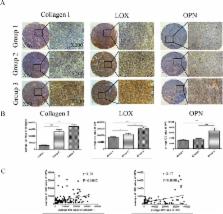
          Increased stromal stiffness is associated with hepatocellular carcinoma (HCC) development and progression. However, the molecular mechanism by which matrix stiffness stimuli modulate HCC progress is largely unknown. In this study, we explored whether matrix stiffness-mediated effects on osteopontin (OPN) expression occur in HCC cells. We used a previously reported in vitro culture system with tunable matrix stiffness and found that OPN expression was remarkably upregulated in HCC cells with increasing matrix stiffness. Furthermore, the phosphorylation level of GSK3β and the expression of nuclear β-catenin were also elevated, indicating that GSK3β/β-catenin pathway might be involved in OPN regulation. Knock-down analysis of integrin β1 showed that OPN expression and p-GSK3β level were downregulated in HCC cells grown on high stiffness substrate compared with controls. Simultaneously, inhibition of GSK-3β led to accumulation of β-catenin in the cytoplasm and its enhanced nuclear translocation, further triggered the rescue of OPN expression, suggesting that the integrin β1/GSK-3β/β-catenin pathway is specifically activated for matrix stiffness-mediated OPN upregulation in HCC cells. Tissue microarray analysis confirmed that OPN expression was positively correlated with the expression of LOX and COL1. Taken together, high matrix stiffness upregulated OPN expression in HCC cells via the integrin β1/GSK-3β/β-catenin signaling pathway. It highlights a new insight into a pathway involving physical mechanical signal and biochemical signal molecules which contributes to OPN expression in HCC cells.

          Related collections

          Most cited references48

          • Record: found
          • Abstract: found
          • Article: not found

          Effects of substrate stiffness on cell morphology, cytoskeletal structure, and adhesion.

          The morphology and cytoskeletal structure of fibroblasts, endothelial cells, and neutrophils are documented for cells cultured on surfaces with stiffness ranging from 2 to 55,000 Pa that have been laminated with fibronectin or collagen as adhesive ligand. When grown in sparse culture with no cell-cell contacts, fibroblasts and endothelial cells show an abrupt change in spread area that occurs at a stiffness range around 3,000 Pa. No actin stress fibers are seen in fibroblasts on soft surfaces, and the appearance of stress fibers is abrupt and complete at a stiffness range coincident with that at which they spread. Upregulation of alpha5 integrin also occurs in the same stiffness range, but exogenous expression of alpha5 integrin is not sufficient to cause cell spreading on soft surfaces. Neutrophils, in contrast, show no dependence of either resting shape or ability to spread after activation when cultured on surfaces as soft as 2 Pa compared to glass. The shape and cytoskeletal differences evident in single cells on soft compared to hard substrates are eliminated when fibroblasts or endothelial cells make cell-cell contact. These results support the hypothesis that mechanical factors impact different cell types in fundamentally different ways, and can trigger specific changes similar to those stimulated by soluble ligands. Copyright 2004 Wiley-Liss, Inc.
            Bookmark
            • Record: found
            • Abstract: found
            • Article: not found

            Matrix stiffness modulates proliferation, chemotherapeutic response, and dormancy in hepatocellular carcinoma cells.

            There is increasing evidence that the physical environment is a critical mediator of tumor behavior. Hepatocellular carcinoma (HCC) develops within an altered biomechanical environment, and increasing matrix stiffness is a strong predictor of HCC development. The aim of this study was to establish whether changes in matrix stiffness, which are characteristic of inflammation and fibrosis, regulate HCC cell proliferation and chemotherapeutic response. Using an in vitro system of "mechanically tunable" matrix-coated polyacrylamide gels, matrix stiffness was modeled across a pathophysiologically relevant range, corresponding to values encountered in normal and fibrotic livers. Increasing matrix stiffness was found to promote HCC cell proliferation. The proliferative index (assessed by Ki67 staining) of Huh7 and HepG2 cells was 2.7-fold and 12.2-fold higher, respectively, when the cells were cultured on stiff (12 kPa) versus soft (1 kPa) supports. This was associated with stiffness-dependent regulation of basal and hepatocyte growth factor-stimulated mitogenic signaling through extracellular signal-regulated kinase, protein kinase B (PKB/Akt), and signal transducer and activator of transcription 3. β1-Integrin and focal adhesion kinase were found to modulate stiffness-dependent HCC cell proliferation. Following treatment with cisplatin, we observed reduced apoptosis in HCC cells cultured on stiff versus soft (physiological) supports. Interestingly, however, surviving cells from soft supports had significantly higher clonogenic capacity than surviving cells from a stiff microenvironment. This was associated with enhanced expression of cancer stem cell markers, including clusters of differentiation 44 (CD44), CD133, c-kit, cysteine-X-cysteine receptor 4, octamer-4 (CXCR4), and NANOG. Increasing matrix stiffness promotes proliferation and chemotherapeutic resistance, whereas a soft environment induces reversible cellular dormancy and stem cell characteristics in HCC. This has implications for both the treatment of primary HCC and the prevention of tumor outgrowth from disseminated tumor cells. (HEPATOLOGY 2011;). Copyright © 2011 American Association for the Study of Liver Diseases.
              Bookmark
              • Record: found
              • Abstract: found
              • Article: not found

              Regulation of Wnt signaling during adipogenesis.

              We have identified Wnt10b as a potent inhibitor of adipogenesis that must be suppressed for preadipocytes to differentiate in vitro. Here, we demonstrate that a specific inhibitor of glycogen synthase kinase 3, CHIR 99021, mimics Wnt signaling in preadipocytes. CHIR 99021 stabilizes free cytosolic beta-catenin and inhibits adipogenesis by blocking induction of CCAAT/enhancer-binding protein alpha and peroxisome proliferator-activated receptor gamma. Preadipocyte differentiation is inhibited when 3T3-L1 cells are exposed to CHIR 99021 for any 24 h period during the first 3 days of adipogenesis. Consistent with this time frame of inhibition, expression of Wnt10b mRNA is suppressed upon induction of differentiation, with a 50% decline by 6 h and complete inhibition by 36 h. Of the agents used to induce differentiation, exposure of 3T3-L1 cells to methyl-isobutylxanthine or cAMP is sufficient to suppress expression of Wnt10b mRNA. Inhibition of adipogenesis by Wnt10b is likely mediated by Wnt receptors, Frizzled 1, 2, and/or 5, and co-receptors low density lipoprotein receptor-related proteins 5 and 6. These receptors, like Wnt10b, are highly expressed in preadipocytes and stromal vascular cells. Finally, we demonstrate that disruption of extracellular Wnt signaling by expression of secreted Frizzled related proteins causes spontaneous adipocyte conversion.
                Bookmark

                Author and article information

                Contributors
                Role: Editor
                Journal
                PLoS One
                PLoS ONE
                plos
                plosone
                PLoS ONE
                Public Library of Science (San Francisco, CA USA )
                1932-6203
                17 August 2015
                2015
                : 10
                : 8
                : e0134243
                Affiliations
                [1 ]Liver Cancer Institute, Zhongshan Hospital, Fudan University & Key Laboratory of Carcinogenesis and Cancer Invasion, Ministry of Education, 136 Xue Yuan Road, Shanghai, 200032, PR China
                [2 ]Department of Interventional Radiology, Shanghai Cancer Center, Fudan University, Shanghai, 200032, PR China
                [3 ]Department of Urology, Zhongshan Hospital, Fudan University, Shanghai, 200032, PR China
                [4 ]Department of Oncology, Zhongshan Hospital Subdivision, Fudan University, Shanghai, 200052, PR China
                The University of Hong Kong, HONG KONG
                Author notes

                Competing Interests: The authors have declared that no competing interests exist.

                Conceived and designed the experiments: YY JFC ZGR. Performed the experiments: YY QDZ YYD. Analyzed the data: YY CH. Contributed reagents/materials/analysis tools: QDZ YYD Yaohui Wang ZMW LZ TCX XYX Yanhong Wang RXC. Wrote the paper: YY JFC ZGR.

                Article
                PONE-D-14-56735
                10.1371/journal.pone.0134243
                4539226
                26280346
                7b65e3fe-9334-433d-81a8-dcd1d7361ec6
                Copyright @ 2015

                This is an open access article distributed under the terms of the Creative Commons Attribution License, which permits unrestricted use, distribution, and reproduction in any medium, provided the original author and source are credited

                History
                : 12 January 2015
                : 7 July 2015
                Page count
                Figures: 5, Tables: 0, Pages: 13
                Funding
                This study was sponsored by grants from the National Natural Science Foundation of China (Nos. 81071902, 81272583) and the Shanghai Science and Technology Programme (11JC1402100).
                Categories
                Research Article
                Custom metadata
                All relevant data are within the paper and its Supporting Information files.

                Uncategorized
                Uncategorized

                Comments

                Comment on this article