15
views
0
recommends
+1 Recommend
0 collections
    0
    shares
      • Record: found
      • Abstract: found
      • Article: found
      Is Open Access

      Oestrogen and parathyroid hormone alleviate lumbar intervertebral disc degeneration in ovariectomized rats and enhance Wnt/β-catenin pathway activity

      research-article

      Read this article at

      Bookmark
          There is no author summary for this article yet. Authors can add summaries to their articles on ScienceOpen to make them more accessible to a non-specialist audience.

          Abstract

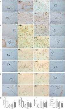
          To investigate the mitigation effect and mechanism of oestrogen and PTH on disc degeneration in rats after ovariectomy, as well as on Wnt/β-catenin pathway activity, thirty 3-month-old rats were ovariectomized and divided into three groups. Ten additional rats were used as controls. Eight weeks later, the rats were administered oestrogen or PTH for 12 weeks, and then discs were collected for tests. Results showed that nucleus pulposus cells in the Sham group were mostly notochord cells, while in the OVX group, cells gradually developed into chondrocyte-like cells. Oestrogen or PTH could partly recover the notochord cell number. After ovariectomy, the endplate roughened and endplate porosity decreased. After oestrogen or PTH treatment, the smoothness and porosity of endplate recovered. Compared with the Sham group, Aggrecan, Col2a and Wnt/β-catenin pathway expression in OVX group decreased, and either oestrogen or PTH treatment improved their expression. The biomechanical properties of intervertebral disc significantly changed after ovariectomy, and oestrogen or PTH treatment partly recovered them. Disc degeneration occurred with low oestrogen, and the underlying mechanisms involve nutrition supply disorders, cell type changes and decreased Wnt/β-catenin pathway activity. Oestrogen and PTH can retard disc degeneration in OVX rats and enhance Wnt/β-catenin pathway activity in nucleus pulposus.

          Related collections

          Most cited references48

          • Record: found
          • Abstract: found
          • Article: not found

          Nutrition of the intervertebral disc.

          A review of the literature on disc nutrition. To summarize the information on disc nutrition in relation to disc degeneration. The disc is avascular, and the disc cells depend on diffusion from blood vessels at the disc's margins to supply the nutrients essential for cellular activity and viability and to remove metabolic wastes such as lactic acid. The nutrient supply can fail due to changes in blood supply, sclerosis of the subchondral bone or endplate calcification, all of which can block transport from blood supply to the disc or due to changes in cellular demand. A review of the studies on disc blood supply, solute transport, studies of solute transport in animal and human disc in vitro, and of theoretical modeling studies that have examined factors affecting disc nutrition. Small nutrients such as oxygen and glucose are supplied to the disc's cells virtually entirely by diffusion; convective transport, arising from load-induced fluid movement in and out of the disc, has virtually no direct influence on transport of these nutrients. Consequently, there are steep concentration gradients of oxygen, glucose, and lactic acid across the disc; oxygen and glucose concentrations are lowest in the center of the nucleus where lactic acid concentrations are greatest. The actual levels of concentration depend on the balance between diffusive transport and cellular demand and can fall to critical levels if the endplate calcifies or nutritional demand increases. Loss of nutrient supply can lead to cell death, loss of matrix production, and increase in matrix degradation and hence to disc degeneration.
            Bookmark
            • Record: found
            • Abstract: found
            • Article: not found

            Histology and pathology of the human intervertebral disc.

            The intervertebral disc is a highly organized matrix laid down by relatively few cells in a specific manner. The central gelatinous nucleus pulposus is contained within the more collagenous anulus fibrosus laterally and the cartilage end plates inferiorly and superiorly. The anulus consists of concentric rings or lamellae, with fibers in the outer lamellae continuing into the longitudinal ligaments and vertebral bodies. This arrangement allows the discs to facilitate movement and flexibility within what would be an otherwise rigid spine. At birth, the human disc has some vascular supply within both the cartilage end plates and the anulus fibrosus, but these vessels soon recede, leaving the disc with little direct blood supply in the healthy adult. With increasing age, water is lost from the matrix, and the proteoglycan content also changes and diminishes. The disc-particularly the nucleus-becomes less gelatinous and more fibrous, and cracks and fissures eventually form. More blood vessels begin to grow into the disc from the outer areas of the anulus. There is an increase in cell proliferation and formation of cell clusters as well as an increase in cell death. The cartilage end plate undergoes thinning, altered cell density, formation of fissures, and sclerosis of the subchondral bone. These changes are similar to those seen in degenerative disc disease, causing discussion as to whether aging and degeneration are separate processes or the same process occurring over a different timescale. Additional disorders involving the intervertebral disc can demonstrate other changes in morphology. Discs from patients with spinal deformities such as scoliosis have ectopic calcification in the cartilage end plate and sometimes in the disc itself. Cells in these discs and cells from patients with spondylolisthesis have been found to have very long cell processes. Cells in herniated discs appear to have a higher degree of cellular senescence than cells in nonherniated discs and produce a greater abundance of matrix metalloproteinases. The role that abnormalities play in the etiopathogenesis of different disorders is not always clear. Disorders may be caused by a genetic predisposition or a tissue response to an insult or altered mechanical environment. Whatever the initial cause, a change in the morphology of the tissue is likely to alter the physiologic and mechanical functioning of the tissue.
              Bookmark
              • Record: found
              • Abstract: found
              • Article: not found

              Vertebral bone mineral density, marrow perfusion, and fat content in healthy men and men with osteoporosis: dynamic contrast-enhanced MR imaging and MR spectroscopy.

              To prospectively use hydrogen 1 (1H) magnetic resonance (MR) spectroscopy and dynamic contrast material-enhanced MR imaging to measure vertebral body marrow fat content and bone marrow perfusion in older men with varying bone mineral densities as documented with dual x-ray absorptiometry (DXA). This study had institutional review board approval, and all participants provided informed consent. DXA, 1H MR spectroscopy, and dynamic contrast-enhanced MR imaging of the lumbar spine were performed in 90 men (mean age, 73 years; range, 67-101 years). Vertebral marrow fat content and perfusion (maximum enhancement and enhancement slope) were compared for subject groups with differing bone densities (normal, osteopenic, and osteoporotic). The t test was used for comparisons between groups, and the Pearson test was used to determine correlation between marrow fat content and perfusion indexes. Eight subjects were excluded, yielding a final cohort of 82 subjects (mean age, 73 years; range, 67-101 years) that included 42 subjects with normal bone density (mean T score, 0.8 +/- 1.1 [standard deviation]), 23 subjects with osteopenia (mean T score, -1.6 +/- 0.4), and 17 subjects with osteoporosis (mean T score, -3.2 +/- 0.5). Vertebral marrow fat content was significantly increased in subjects with osteoporosis (mean fat content, 58.23% +/- 7.8) (P = .002) or osteopenia (mean fat content, 55.68% +/- 10.2) (P = .034) compared with that in subjects with normal bone density (50.45% +/- 8.7). Vertebral marrow perfusion indexes were significantly decreased in osteoporotic subjects (mean enhancement slope, 0.78%/sec +/- 0.3) compared with those in osteopenic subjects (mean enhancement slope, 1.15%/sec +/- 0.6) (P = .007) and those in subjects with normal bone density (mean enhancement slope, 1.48%/sec +/- 0.7) (P < .001). Subjects with osteoporosis have decreased vertebral marrow perfusion and increased marrow fat compared with these parameters in subjects with osteopenia. Similarly, subjects with osteopenia have decreased vertebral marrow perfusion and increased marrow fat compared with these parameters in subjects with normal bone density.
                Bookmark

                Author and article information

                Journal
                Sci Rep
                Sci Rep
                Scientific Reports
                Nature Publishing Group
                2045-2322
                09 June 2016
                2016
                : 6
                : 27521
                Affiliations
                [1 ]Department of Orthopedics Institute, Tianjin Hospital , 406, Jiefang Nan Street, Hexi District, Tianjin 300211, China
                [2 ]Biomechanics Labs of Orthopedic Research Institute, Tianjin Hospital Heping Branch , 122, MuNan Street, Heping District, Tianjin 300050, China
                [3 ]Tianjin Medical University , No. 22, Qixiangtai Road, Heping District, Tianjin 300070, China
                Author notes
                [*]

                These authors contributed equally to this work.

                Article
                srep27521
                10.1038/srep27521
                4899752
                27279629
                7bc8825e-8491-44db-87a3-9ad867d53ae1
                Copyright © 2016, Macmillan Publishers Limited

                This work is licensed under a Creative Commons Attribution 4.0 International License. The images or other third party material in this article are included in the article’s Creative Commons license, unless indicated otherwise in the credit line; if the material is not included under the Creative Commons license, users will need to obtain permission from the license holder to reproduce the material. To view a copy of this license, visit http://creativecommons.org/licenses/by/4.0/

                History
                : 25 November 2015
                : 09 May 2016
                Categories
                Article

                Uncategorized
                Uncategorized

                Comments

                Comment on this article