18
views
0
recommends
+1 Recommend
0 collections
    0
    shares
      • Record: found
      • Abstract: found
      • Article: found
      Is Open Access

      Correlation between Patent Foramen Ovale, Cerebral “Lesions” and Neuropsychometric Testing in Experienced Sports Divers: Does Diving Damage the Brain?

      research-article

      Read this article at

      Bookmark
          There is no author summary for this article yet. Authors can add summaries to their articles on ScienceOpen to make them more accessible to a non-specialist audience.

          Abstract

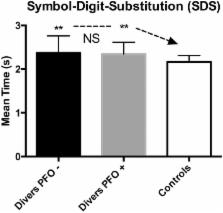
          SCUBA diving exposes divers to decompression sickness (DCS). There has been considerable debate whether divers with a Patent Foramen Ovale of the heart have a higher risk of DCS because of the possible right-to-left shunt of venous decompression bubbles into the arterial circulation. Symptomatic neurological DCS has been shown to cause permanent damage to brain and spinal cord tissue; it has been suggested that divers with PFO may be at higher risk of developing subclinical brain lesions because of repeated asymptomatic embolization of decompression-induced nitrogen bubbles. These studies however suffer from several methodological flaws, including self-selection bias. We recruited 200 volunteer divers from a recreational diving population who had never suffered from DCS; we then randomly selected 50 of those for further investigation. The selected divers underwent brain Magnetic Resonance Imaging to detect asymptomatic brain lesions, contrast trans-oesophageal echocardiography for PFO, and extensive neuro-psychometric testing. Neuro-psychometry results were compared with a control group of normal subjects and a separate control group for subjects exposed to neurotoxic solvents. Forty two divers underwent all the tests and are included in this report. Grade 2 Patent Foramen Ovale was found in 16 (38%) of the divers; brain Unidentified Bright Objects (UBO's) were found in 5 (11.9%). There was no association between PFO and the presence of UBO's ( P = 0.693) or their size ( p = 0.5) in divers. Neuropsychometric testing in divers was significantly worse from controls in two tests, Digit Span Backwards (DSB; p < 0.05) and Symbol-Digit-Substitution (SDS; p < 0.01). Compared to subjects exposed to neurotoxic solvents, divers scored similar on DSB and SDS tests, but significantly better on the Simple Reaction Time (REA) and Hand-Eye Coordination (EYE) tests. There was no correlation between PFO, number of UBO's and any of the neuro-psychometric tests. We conclude that for uneventful recreational diving, PFO does not appear to influence the presence of UBO's. Diving by itself seems to cause some decrease of short-term memory and higher cognitive function, including visual-motor skills; this resembles some of the effects of nitrogen narcosis and we suggest that this may be a prolonged effect of diving.

          Related collections

          Most cited references59

          • Record: found
          • Abstract: found
          • Article: not found

          Incidence and size of patent foramen ovale during the first 10 decades of life: an autopsy study of 965 normal hearts.

          The incidence and size of the patent foramen ovale were studied in 965 autopsy specimens of human hearts, which were from subjects who were evenly distributed by sex and age. Neither incidence nor size of the defect was significantly different between male and female subjects. The overall incidence was 27.3%, but it progressively declined with increasing age from 34.3% during the first three decades of life to 25.4% during the 4th through 8th decades and to 20.2% during the 9th and 10th decades. Among the 263 specimens that exhibited patency in our study, the foramen ovale ranged from 1 to 19 mm in maximal potential diameter (mean, 4.9 mm). In 98% of these cases, the foramen ovale was 1 to 10 mm in diameter. The size tended to increase with increasing age, from a mean of 3.4 mm in the first decade to 5.8 mm in the 10th decade of life.
            Bookmark
            • Record: found
            • Abstract: found
            • Article: not found

            Effects of frontal lobe damage on interference effects in working memory.

            Working memory is hypothesized to comprise a collection of distinct components or processes, each of which may have a unique neural substrate. Recent neuroimaging studies have isolated a region of the left inferior frontal gyrus that appears to be related specifically to one such component: resolving interference from previous items in working memory. In the present study, we examined working memory in patients with unilateral frontal lobe lesions by using a modified version of an item recognition task in which interference from previous trials was manipulated. In particular, we focused on patient R.C., whose lesion uniquely impinged on the region identified in the neuroimaging studies of interference effects. We measured baseline working memory performance and interference effects in R.C. and other frontal patients and in age-matched control subjects and young control subjects. Comparisons of each of these groups supported the following conclusions. Normal aging is associated with changes to both working memory and interference effects. Patients with frontal damage exhibited further declines in working memory but normal interference effects, with the exception of R.C., who exhibited a pronounced interference effect on both response time and accuracy. We propose that the left inferior frontal gyrus subserves a general, nonmnemonic function of selecting relevant information in the face of competing alternatives and that this function may be required by some working memory tasks.
              Bookmark
              • Record: found
              • Abstract: found
              • Article: not found

              Cohort study of multiple brain lesions in sport divers: role of a patent foramen ovale.

              To investigate the role of a patient foramen ovale in the pathogenesis of multiple brain lesions acquired by sport divers in the absence of reported decompression symptoms. Prospective double blind cohort study. Diving clubs around Heidelberg and departments of neuroradiology and neurology. 87 sport divers with a minimum of 160 scuba dives (dives with self contained underwater breathing apparatus). Presence of multiple brain lesions visualised by cranial magnetic resonance imaging and presence and size of patent foramen ovale as documented by echocontrast transcranial Doppler ultrasonography. 25 subjects were found to have a right-to-left shunt, 13 with a patent foramen ovale of high haemodynamic relevance. A total of 41 brain lesions were detected in 11 divers. There were seven brain lesions in seven divers without a right-to-left shunt and 34 lesions in four divers with a right-to-left shunt. Multiple brain lesions occurred exclusively in three divers with a large patent foramen ovale (P = 0.004). Multiple brain lesions in sport divers were associated with presence of a large patent foramen ovale. This association suggests paradoxical gas embolism as the pathological mechanism. A patent foramen ovale of high haemodynamic relevance seems to be an important risk factor for developing multiple brain lesions in sport divers.
                Bookmark

                Author and article information

                Contributors
                Journal
                Front Psychol
                Front Psychol
                Front. Psychol.
                Frontiers in Psychology
                Frontiers Media S.A.
                1664-1078
                11 May 2016
                2016
                : 7
                : 696
                Affiliations
                [1] 1DAN Europe Research Division Brussels, Belgium
                [2] 2Faculté des Sciences de la Motricité, Université Libre de Bruxelles Brussels, Belgium
                [3] 3Motor Sciences and Physiotherapy, Environmental and Occupational (Integrative) Physiology, Haute Ecole Paul Henri Spaak Brussels, Belgium
                [4] 4Center for Hyperbaric Oxygen Therapy, Military Hospital Queen Astrid Brussels, Belgium
                Author notes

                Edited by: Guy Cheron, Université Libre de Bruxelles, Belgium

                Reviewed by: Patrice Jissendi Tchofo, Saint-Pierre Universitiy Hospital, Belgium; Jacek Kot, Medical University of Gdansk, Poland

                *Correspondence: Costantino Balestra costantinobalestra@ 123456gmail.com

                This article was submitted to Movement Science and Sport Psychology, a section of the journal Frontiers in Psychology

                †These authors have contributed equally to this work and co-first authors.

                Article
                10.3389/fpsyg.2016.00696
                4863080
                27242609
                7bf41c0a-bb87-4cae-993c-156dfbc80369
                Copyright © 2016 Balestra and Germonpré.

                This is an open-access article distributed under the terms of the Creative Commons Attribution License (CC BY). The use, distribution or reproduction in other forums is permitted, provided the original author(s) or licensor are credited and that the original publication in this journal is cited, in accordance with accepted academic practice. No use, distribution or reproduction is permitted which does not comply with these terms.

                History
                : 09 September 2015
                : 26 April 2016
                Page count
                Figures: 4, Tables: 0, Equations: 0, References: 64, Pages: 8, Words: 7187
                Categories
                Psychology
                Original Research

                Clinical Psychology & Psychiatry
                adverse effects,scuba diving,pfo,embolism,long term effects
                Clinical Psychology & Psychiatry
                adverse effects, scuba diving, pfo, embolism, long term effects

                Comments

                Comment on this article