75
views
0
recommends
+1 Recommend
0 collections
    0
    shares
      • Record: found
      • Abstract: found
      • Article: found
      Is Open Access

      Myostatin Is Elevated in Congenital Heart Disease and After Mechanical Unloading

      research-article

      Read this article at

      Bookmark
          There is no author summary for this article yet. Authors can add summaries to their articles on ScienceOpen to make them more accessible to a non-specialist audience.

          Abstract

          Background

          Myostatin is a negative regulator of skeletal muscle mass whose activity is upregulated in adult heart failure (HF); however, its role in congenital heart disease (CHD) is unknown.

          Methods

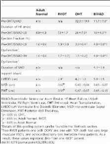
          We studied myostatin and IGF-1 expression via Western blot in cardiac tissue at varying degrees of myocardial dysfunction and after biventricular support in CHD by collecting myocardial biopsies from four patient cohorts: A) adult subjects with no known cardiopulmonary disease (left ventricle, LV), (Adult Normal), (n = 5); B) pediatric subjects undergoing congenital cardiac surgery with normal RV size and function (right ventricular outflow tract, RVOT), (n = 3); C) pediatric subjects with worsening but hemodynamically stable LV failure [LV and right ventricle (LV, RV,)] with biopsy collected at the time of orthotopic heart transplant (OHT), (n = 7); and D) pediatric subjects with decompensated bi-ventricular failure on BiVAD support with biopsy collected at OHT (LV, RV, BiVAD), (n = 3).

          Results

          The duration of HF was longest in OHT patients compared to BIVAD. The duration of BiVAD support was 4.3±1.9 days. Myostatin expression was significantly increased in LV-OHT compared to RV-OHT and RVOT, and was increased more than double in decompensated biventricular HF (BiVAD) compared to both OHT and RVOT. An increased myostatin/IGF-1 ratio was associated with ventricular dysfunction.

          Conclusions

          Myostatin expression in increased in CHD, and the myostatin/IGF-1 ratio increases as ventricular function deteriorates. Future investigation is necessary to determine if restoration of the physiologic myostatin/IGF-1 ratio has therapeutic potential in HF.

          Related collections

          Most cited references27

          • Record: found
          • Abstract: found
          • Article: not found

          Myostatin decreases with aerobic exercise and associates with insulin resistance.

          There is mounting evidence that skeletal muscle produces and secretes biologically active proteins or "myokines" that facilitate metabolic cross talk between organ systems. The increased expression of myostatin, a secreted anabolic inhibitor of muscle growth and development, has been associated with obesity and insulin resistance. Despite these intriguing findings, there have been few studies linking myostatin and insulin resistance. To explore this relationship in more detail, we quantified myostatin protein in muscle and plasma from 10 insulin-resistant, middle-aged (53.1 ± 5.5 yr) men before and after 6 months of moderate aerobic exercise training (1200 kcal·wk−¹ at 40%-55% VO2peak). To establish a cause-effect relationship, we also injected C57/Bl6 male mice with high physiological levels of recombinant myostatin protein. Myostatin protein levels were shown to decrease in muscle (37%, P = 0.042, n = 10) and matching plasma samples (from 28.7 ng·mL−¹ pretraining to 22.8 ng·mL−¹ posttraining, P = 0.003, n = 9) with aerobic exercise. Furthermore, the strong correlation between plasma myostatin levels and insulin sensitivity (R² = 0.82, P < 0.001, n = 9) suggested a cause-effect relationship that was subsequently confirmed by inducing insulin resistance in myostatin-injected mice. A modest increase (44%) in plasma myostatin levels was also associated with significant reductions in the insulin-stimulated phosphorylation of Akt (Thr308) in both muscle and liver of myostatin-treated animals. These findings indicate that both muscle and plasma myostatin protein levels are regulated by aerobic exercise and, furthermore, that myostatin is in the causal pathway of acquired insulin resistance with physical inactivity.
            Bookmark
            • Record: found
            • Abstract: found
            • Article: not found

            Genetic deletion of myostatin from the heart prevents skeletal muscle atrophy in heart failure.

            Cardiac cachexia is characterized by an exaggerated loss of skeletal muscle, weakness, and exercise intolerance, although the cause of these effects remains unknown. Here, we hypothesized that the heart functions as an endocrine organ in promoting systemic cachexia by secreting peptide factors such as myostatin. Myostatin is a cytokine of the transforming growth factor-beta superfamily that is known to control muscle wasting. We used a Cre/loxP system to ablate myostatin (Mstn gene) expression in a cell type-specific manner. As expected, elimination of Mstn selectively in skeletal muscle with a myosin light chain 1f (MLC1f)-cre allele induced robust hypertrophy in all skeletal muscle. However, heart-specific deletion of Mstn with an Nkx2.5-cre allele did not alter baseline heart size or secondarily affect skeletal muscle size, but the characteristic wasting and atrophy of skeletal muscle that typify heart failure were not observed in these heart-specific null mice, indicating that myocardial myostatin expression controls muscle atrophy in heart failure. Indeed, myostatin levels in the plasma were significantly increased in wild-type mice subjected to pressure overload-induced cardiac hypertrophy but not in Mstn heart-specific deleted mice. Moreover, cardiac-specific overexpression of myostatin, which increased circulating levels of myostatin by 3- to 4-fold, caused a reduction in weight of the quadriceps, gastrocnemius, soleus, and even the heart itself. Finally, to investigate myostatin as a potential therapeutic target for the treatment of muscle wasting in heart failure, we infused a myostatin blocking antibody (JA-16), which promoted greater maintenance of muscle mass in heart failure. Myostatin released from cardiomyocytes induces skeletal muscle wasting in heart failure. Targeted inhibition of myostatin in cardiac cachexia might be a therapeutic option in the future.
              Bookmark
              • Record: found
              • Abstract: found
              • Article: not found

              Myostatin, a transforming growth factor-beta superfamily member, is expressed in heart muscle and is upregulated in cardiomyocytes after infarct.

              Myostatin is a secreted growth and differentiating factor (GDF-8) that belongs to the transforming growth factor-beta (TGF-beta) superfamily. Targeted disruption of the myostatin gene in mice and a mutation in the third exon of the myostatin gene in double-muscled Belgian Blue cattle breed result in skeletal muscle hyperplasia. Hence, myostatin has been shown to be involved in the regulation of skeletal muscle mass in both mice and cattle. Previous published reports utilizing Northern hybridization had shown that myostatin expression was seen exclusively in skeletal muscle. A significantly lower level of myostatin mRNA was also reported in adipose tissue. Using a sensitive reverse transcription-polymerase chain reaction (RT-PCR) technique and Western blotting with anti-myostatin antibodies, we show that myostatin mRNA and protein are not restricted to skeletal muscle. We also show that myostatin expression is detected in the muscle of both fetal and adult hearts. Sequence analysis reveals that the Belgian Blue heart myostatin cDNA sequence contains an 11 nucleotide deletion in the third exon that causes a frameshift that eliminates virtually all of the mature, active region of the protein. Anti-myostatin immunostaining on heart sections also demonstrates that myostatin protein is localized in Purkinje fibers and cardiomyocytes in heart tissue. Furthermore, following myocardial infarction, myostatin expression is upregulated in the cardiomyocytes surrounding the infarct area. Given that myostatin is expressed in fetal and adult hearts and that myostatin expression is upregulated in cardiomyocytes after the infarction, myostatin could play an important role in cardiac development and physiology.
                Bookmark

                Author and article information

                Contributors
                Role: Editor
                Journal
                PLoS One
                plos
                plosone
                PLoS ONE
                Public Library of Science (San Francisco, USA )
                1932-6203
                2011
                13 September 2011
                : 6
                : 9
                : e23818
                Affiliations
                [1 ]Department of Physiology, University of Pennsylvania School of Medicine, Philadelphia, Pennsylvania, United States of America
                [2 ]Department of Surgery, Division of Cardiothoracic Surgery, College of Physicians and Surgeons, Columbia University, New York, New York, United States of America
                [3 ]Department of Medicine, Division of Cardiology, Albert Einstein College of Medicine, New York, New York, United States of America
                Medical College of Georgia, United States of America
                Author notes

                Conceived and designed the experiments: LB IG SM JY JC HS. Performed the experiments: LB IG. Analyzed the data: LB IG. Contributed reagents/materials/analysis tools: HS. Wrote the paper: LB IG.

                Article
                PONE-D-11-07106
                10.1371/journal.pone.0023818
                3172210
                21931616
                7cd873c9-425b-42e7-9170-d61b54563381
                Bish et al. This is an open-access article distributed under the terms of the Creative Commons Attribution License, which permits unrestricted use, distribution, and reproduction in any medium, provided the original author and source are credited.
                History
                : 22 April 2011
                : 26 July 2011
                Page count
                Pages: 8
                Categories
                Research Article
                Biology
                Biochemistry
                Proteins
                Growth Factors
                Genetics
                Gene Expression
                Molecular Cell Biology
                Cell Growth
                Medicine
                Cardiovascular
                Cardiomyopathies
                Congenital Heart Disease
                Heart Failure
                Pediatric Cardiology
                Surgery
                Cardiovascular Surgery
                Pediatric Surgery

                Uncategorized
                Uncategorized

                Comments

                Comment on this article