45
views
0
recommends
+1 Recommend
0 collections
    0
    shares
      • Record: found
      • Abstract: found
      • Article: found
      Is Open Access

      Escape of Actively Secreting Shigella flexneri from ATG8/LC3-Positive Vacuoles Formed during Cell-To-Cell Spread Is Facilitated by IcsB and VirA

      research-article

      Read this article at

      Bookmark
          There is no author summary for this article yet. Authors can add summaries to their articles on ScienceOpen to make them more accessible to a non-specialist audience.

          ABSTRACT

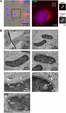
          The enteropathogenic bacterium Shigella flexneri uses a type 3 secretion apparatus (T3SA) to transfer proteins dubbed translocators and effectors inside host cells, inducing bacterial uptake and subsequent lysis of the entry vacuole. Once in the cytoplasm, the outer membrane protein IcsA induces actin polymerization, enabling cytoplasmic movement and cell-to-cell spread of bacteria. During this infectious process, S. flexneri is targeted by ATG8/LC3. The effector IcsB was proposed to inhibit LC3 recruitment by masking a region of IcsA recognized by the autophagy pathway component ATG5. The effector VirA, a GTPase-activating protein (GAP) for Rab1, was also shown to prevent LC3 recruitment. However, the context of LC3 recruitment around S. flexneri is not fully understood. Here, we show that LC3 is recruited specifically around secreting bacteria that are still present in vacuoles formed during entry and cell-to-cell spread. While LC3 recruitment occurs around a small proportion of intracellular wild-type bacteria, the icsB, virA, and icsB virA mutants display incremental defaults in escape from LC3-positive vacuoles formed during cell-to-cell spread. Our results indicate that IcsB and VirA act synergistically to allow bacteria to escape from LC3-positive vacuoles by acting at or in the immediate vicinity of the vacuole membrane(s). We also demonstrate that LC3 is recruited around bacteria still present in the single-membrane entry vacuole, in a manner akin to that seen with LC3-associated phagocytosis. Our results indicate that LC3 recruitment occurs around bacteria still, or already, in membrane compartments formed during entry and cell-to-cell spread, and not around bacteria free in the cytoplasm.

          IMPORTANCE

          The targeting of S. flexneri by LC3 is a classic example of the targeting of foreign cytoplasmic particles by autophagy (so-called “xenoautophagy”). It is often assumed that LC3 is recruited around bacteria present in the cytoplasm through the formation of canonical double-membrane autophagosomes. Our results indicate that LC3 is recruited around the entry vacuole composed of a single membrane as in the case of LC3-associated phagocytosis. Effectors IcsB and VirA had been implicated in the blocking of LC3 recruitment, but it was not known if they acted on the same or distinct LC3-recruiting pathways. Our results indicate that LC3 is recruited exclusively around bacteria present in vacuoles formed during entry and cell-to-cell spread and that both IcsB and VirA intervene at the latter stage to facilitate bacterial escape. Our report reconciles several findings and may have broad implications for our understanding of the specific targeting of bacterial pathogens by LC3.

          Related collections

          Most cited references31

          • Record: found
          • Abstract: found
          • Article: not found

          The TBK1 adaptor and autophagy receptor NDP52 restricts the proliferation of ubiquitin-coated bacteria.

          Cell-autonomous innate immune responses against bacteria attempting to colonize the cytosol of mammalian cells are incompletely understood. Polyubiquitylated proteins can accumulate on the surface of such bacteria, and bacterial growth is restricted by Tank-binding kinase (TBK1). Here we show that NDP52, not previously known to contribute to innate immunity, recognizes ubiquitin-coated Salmonella enterica in human cells and, by binding the adaptor proteins Nap1 and Sintbad, recruits TBK1. Knockdown of NDP52 and TBK1 facilitated bacterial proliferation and increased the number of cells containing ubiquitin-coated salmonella. NDP52 also recruited LC3, an autophagosomal marker, and knockdown of NDP52 impaired autophagy of salmonella. We conclude that human cells utilize the ubiquitin system and NDP52 to activate autophagy against bacteria attempting to colonize their cytosol.
            Bookmark
            • Record: found
            • Abstract: found
            • Article: not found

            Escape of intracellular Shigella from autophagy.

            The degradation of undesirable cellular components or organelles, including invading microbes, by autophagy is crucial for cell survival. Here, Shigella, an invasive bacteria, was found to be able to escape autophagy by secreting IcsB by means of the type III secretion system. Mutant bacteria lacking IcsB were trapped by autophagy during multiplication within the host cells. IcsB did not directly inhibit autophagy. Rather, Shigella VirG, a protein required for intracellular actin-based motility, induced autophagy by binding to the autophagy protein, Atg5. In nonmutant Shigella, this binding is competitively inhibited by IcsB binding to VirG.
              Bookmark
              • Record: found
              • Abstract: found
              • Article: not found

              Bacterial invasion: the paradigms of enteroinvasive pathogens.

              Invasive bacteria actively induce their own uptake by phagocytosis in normally nonphagocytic cells and then either establish a protected niche within which they survive and replicate, or disseminate from cell to cell by means of an actin-based motility process. The mechanisms underlying bacterial entry, phagosome maturation, and dissemination reveal common strategies as well as unique tactics evolved by individual species to establish infection.
                Bookmark

                Author and article information

                Journal
                mBio
                MBio
                mbio
                mbio
                mBio
                mBio
                American Society of Microbiology (1752 N St., N.W., Washington, DC )
                2150-7511
                26 May 2015
                May-Jun 2015
                : 6
                : 3
                : e02567-14
                Affiliations
                [ a ]Institut Pasteur, Unité de Pathogénie Microbienne Moléculaire, Paris, France
                [ b ]INSERM, U1202, Paris, France
                [ c ]Institut Pasteur, Ultrapole, Paris, France
                [ d ]Collège de France, Chaire de Microbiologie et Maladies Infectieuses, Paris, France
                Author notes
                Address correspondence to François-Xavier Campbell-Valois, fx.campbell.valois@ 123456gmail.com .

                Editor Arturo Casadevall, Johns Hopkins Bloomberg School of Public Health

                This article is a direct contribution from a Fellow of the American Academy of Microbiology.

                Article
                mBio02567-14
                10.1128/mBio.02567-14
                4447254
                26015503
                7fd8d3d0-9c40-44f2-aab3-428d942ec598
                Copyright © 2015 Campbell-Valois et al.

                This is an open-access article distributed under the terms of the Creative Commons Attribution-Noncommercial-ShareAlike 3.0 Unported license, which permits unrestricted noncommercial use, distribution, and reproduction in any medium, provided the original author and source are credited.

                History
                : 19 March 2015
                : 23 April 2015
                Page count
                supplementary-material: 10, Figures: 5, Tables: 0, Equations: 0, References: 46, Pages: 11, Words: 10579
                Categories
                Research Article
                Custom metadata
                May/June 2015

                Life sciences
                Life sciences

                Comments

                Comment on this article