18
views
0
recommends
+1 Recommend
0 collections
    0
    shares
      • Record: found
      • Abstract: found
      • Article: found
      Is Open Access

      Divergent calcium signaling in RBCs from Tropidurus torquatus (Squamata – Tropiduridae) strengthen classification in lizard evolution

      research-article
      1 , 2 ,
      BMC Physiology
      BioMed Central

      Read this article at

      Bookmark
          There is no author summary for this article yet. Authors can add summaries to their articles on ScienceOpen to make them more accessible to a non-specialist audience.

          Abstract

          Background

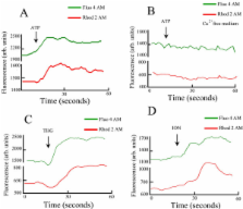
          We have previously reported that a Teiid lizard red blood cells (RBCs) such as Ameiva ameiva and Tupinambis merianae controls intracellular calcium levels by displaying multiple mechanisms. In these cells, calcium stores could be discharged not only by: thapsigargin, but also by the Na +/H + ionophore monensin, K +/H + ionophore nigericin and the H + pump inhibitor bafilomycin as well as ionomycin. Moreover, these lizards possess a P2Y-type purinoceptors that mobilize Ca 2+ from intracellular stores upon ATP addition.

          Results

          Here we report, that RBCs from the tropidurid lizard Tropidurus torquatus store Ca 2+ in endoplasmic reticulum (ER) pool but unlike in the referred Teiidae, these cells do not store calcium in monensin-nigericin sensitive pools. Moreover, mitochondria from T. torquatus RBCs accumulate Ca 2+. Addition of ATP to a calcium-free medium does not increase the [Ca 2+] c levels, however in a calcium medium we observe an increase in cytosolic calcium. This is an indication that purinergic receptors in these cells are P2X-like.

          Conclusion

          T. torquatus RBCs present different mechanisms from Teiid lizard red blood cells (RBCs), for controlling its intracellular calcium levels. At T. torquatus the ion is only stored at endoplasmic reticulum and mitochondria. Moreover activation of purinergic receptor, P2X type, was able to induce an influx of calcium from extracelullar medium. These studies contribute to the understanding of the evolution of calcium homeostasis and signaling in nucleated RBCs.

          Related collections

          Most cited references36

          • Record: found
          • Abstract: found
          • Article: not found

          Microdomains with high Ca2+ close to IP3-sensitive channels that are sensed by neighboring mitochondria.

          Microdomains of high intracellular calcium ion concentration, [Ca2+]i, have been hypothesized to occur in living cells exposed to stimuli that generate inositol 1,4,5-trisphosphate (IP3). Mitochondrially targeted recombinant aequorin was used to show that IP3-induced Ca2+ mobilization from intracellular stores caused increases of mitochondrial Ca2+ concentration, [Ca2+]m, the speed and amplitude of which are not accounted for by the relatively small increases in mean [Ca2+]i. A similar response was obtained by the addition of IP3 to permeabilized cells but not by perfusion of cells with Ca2+ at concentrations similar to those measured in intact cells. It is concluded that in vivo, domains of high [Ca2+]i are transiently generated close to IP3-gated channels and sensed by nearby mitochondria; this may provide an efficient mechanism for optimizing mitochondrial activity upon cell stimulation.
            Bookmark
            • Record: found
            • Abstract: found
            • Article: not found

            Rapid changes of mitochondrial Ca2+ revealed by specifically targeted recombinant aequorin.

            Introduction of Ca2+ indicators (photoproteins, fluorescent dyes) that can be trapped in the cytosolic compartment of living cells has yielded major advances in our knowledge of Ca2+ homeostasis. Ca2+ however regulates functions not only in the cytosol but also within various organelles where indicators have not yet been specifically targeted. Here we present a novel procedure by which the free Ca2+ concentration of mitochondria, [Ca2+]m, can be monitored continuously at rest and during stimulation. The complementary DNA for the Ca2+ sensitive photoprotein aequorin was fused in frame with that encoding a mitochondrial presequence. The hybrid cDNA was transfected into bovine endothelial cells and stable clones were obtained expressing variable amounts of mitochondrially targeted apoaequorin. The functional photoprotein could be reconstituted in intact cells by incubation with purified coelenterazine and [Ca2+]m could thus be monitored in situ. This allowed the unprecedented direct demonstration that agonist-stimulated elevations of cytosolic free Ca2+, [Ca2+]i, (measured in parallel with Fura-2) evoke rapid and transient increases of [Ca2+]m, which can be prevented by pretreatment with a mitochondrial uncoupler. The possibility of targeting aequorin to cellular organelles not only offers a new and powerful method for studying aspects of Ca2+ homeostasis that up to now could not be directly approached, but might also be used in the future as a tool to report in situ a variety of apparently unrelated phenomena of wide biological interest.
              Bookmark
              • Record: found
              • Abstract: found
              • Article: not found

              Decoding of cytosolic calcium oscillations in the mitochondria.

              Frequency-modulated oscillations of cytosolic Ca2+ ([Ca2+]c) are believed to be important in signal transduction, but it has been difficult to correlate [Ca2+]c oscillations directly with the activity of Ca(2+)-regulated targets. We have studied the control of Ca(2+)-sensitive mitochondrial dehydrogenases (CSMDHs) by monitoring mitochondrial Ca2+ ([Ca2+]m) and the redox state of flavoproteins and pyridine nucleotides simultaneously with [Ca2+]c in single hepatocytes. Oscillations of [Ca2+]c induced by IP3-dependent hormones were efficiently transmitted to the mitochondria as [Ca2+]m oscillations. Each [Ca2+]m spike was sufficient to cause a maximal transient activation of the CSMDHs and [Ca2+]m oscillations at frequencies above 0.5 per minute caused a sustained activation of mitochondrial metabolism. By contrast, sustained [Ca2+]c increases yielded only transient CSMDH activation, and slow or partial [Ca2+]c elevations were ineffective in increasing [Ca2+]m or stimulating CSMDHs. We conclude that the mitochondria are tuned to oscillating [Ca2+]c signals, the frequency of which can control the CSMDHs over the full range of potential activities.
                Bookmark

                Author and article information

                Journal
                BMC Physiol
                BMC Physiology
                BioMed Central (London )
                1472-6793
                2007
                23 August 2007
                : 7
                : 7
                Affiliations
                [1 ]Universidade de São Paulo, Instituto de Ciências Biomédicas, Departamento de Parasitologia, São Paulo, Brazil
                [2 ]Universidade de São Paulo, Instituto de Biociências, Departamento de Fisiologia, São Paulo, Brazil
                Article
                1472-6793-7-7
                10.1186/1472-6793-7-7
                2018699
                17716375
                805d23cb-06b6-4750-aa8d-74a28663f96c
                Copyright © 2007 Beraldo and Garcia; licensee BioMed Central Ltd.

                This is an Open Access article distributed under the terms of the Creative Commons Attribution License ( http://creativecommons.org/licenses/by/2.0), which permits unrestricted use, distribution, and reproduction in any medium, provided the original work is properly cited.

                History
                : 13 December 2006
                : 23 August 2007
                Categories
                Research Article

                Anatomy & Physiology
                Anatomy & Physiology

                Comments

                Comment on this article