8
views
0
recommends
+1 Recommend
0 collections
    0
    shares
      • Record: found
      • Abstract: found
      • Article: found
      Is Open Access

      Comparison of a Distal Tibial Allograft and Scapular Spinal Autograft for Posterior Shoulder Instability With Glenoid Bone Loss

      research-article

      Read this article at

      Bookmark
          There is no author summary for this article yet. Authors can add summaries to their articles on ScienceOpen to make them more accessible to a non-specialist audience.

          Abstract

          Background:

          Posterior glenoid bone deficiency can occur with recurrent glenohumeral instability. Glenoid reconstruction with a distal tibial allograft (DTA) has been reported to successfully restore contact pressures that occur during posterior glenohumeral translation. However, there are concerns regarding the risk of allograft resorption, availability, and costs. Extracapsular reconstruction using a scapular spinal autograft (SSA) has been reported as an alternative technique secondary to its anatomic location relative to the posterior shoulder and preferable autograft properties. There are no known prior biomechanical studies evaluating the scapular spine as an effective extracapsular graft choice.

          Purpose:

          To compare the efficacy of a DTA and SSA in restoring the stability of a glenoid with a large posterior bone defect compared with the intact native glenoid.

          Study Design:

          Controlled laboratory study.

          Methods:

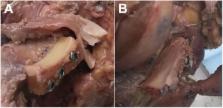
          Ten cadaveric shoulders were tested. With the use of a custom KUKA robot, a 50-N compressive force was applied to the glenohumeral joint. The peak force required to translate the humeral head beyond the glenoid lip posteriorly as well as the lateral displacement that occurred during posterior translation were measured. Testing was performed in 5 conditions: (1) intact glenoid and labrum, (2) simulated reverse Bankart lesion, (3) 12-mm posterior glenoid defect, (4) glenoid defect reconstructed with a fresh DTA, and (5) glenoid defect reconstructed with an SSA.

          Results:

          The mean glenoid width was 30 mm. The mean peak force and lateral displacement decreased significantly with a glenoid defect (0.99 ± 2.3 N and 0.06 ± 0.09 mm, respectively; P < .0001) compared with the intact glenoid (23.00 ± 9.7 N and 1.83 ± 0.70 mm, respectively; P = .0001). There was no significant difference between the peak force after reconstruction of the defect with a DTA (23.00 ± 7.4 N) and SSA (23.00 ± 7.7 N) when compared with the intact glenoid ( P = .9999). There were no significant differences in the peak force between the 2 grafts ( P = .9999). Additionally, both the DTA (1.04 ± 1.09 mm) and the SSA (1.02 ± 1.17 mm) demonstrated no differences in lateral displacement when compared with the intact glenoid ( P = .2336 and .2043, respectively). There was no difference in lateral displacement that occurred between the DTA and SSA ( P = .9999).

          Conclusion:

          Reconstruction of a large posterior glenoid defect with either a DTA or an SSA can effectively restore glenohumeral stability.

          Clinical Relevance:

          This study supports the use of a DTA or SSA in posterior glenoid defect reconstruction. Clinical studies are needed to determine the long-term effects of utilizing such grafts.

          Related collections

          Most cited references17

          • Record: found
          • Abstract: found
          • Article: not found

          The effect of a glenoid defect on anteroinferior stability of the shoulder after Bankart repair: a cadaveric study.

          An osseous defect of the glenoid rim is sometimes caused by multiple recurrent dislocations of the shoulder. It is generally thought that a large defect should be treated with bone-grafting, but there is a lack of consensus with regard to how large a defect must be in order to necessitate this procedure. Some investigators have proposed that a defect must involve at least one-third of the glenoid surface in order to necessitate bone-grafting. However, it is difficult to determine (1) whether a defect involves one-third of the glenoid surface and (2) whether a defect of this size is critical to the stability of the shoulder after a Bankart repair. The purposes of the present study were (1) to create and quantify various sizes of osseous defects of the glenoid and (2) to determine the effect of such defects on the stability and motion of the shoulder after Bankart repair. The glenoids from sixteen dried scapulae were photographed, and the images were scanned into a computer. The average shape of the glenoid was determined on the basis of the scans, and this information was used to design custom templates for the purpose of creating various sizes of osseous defects. Ten fresh-frozen cadaveric shoulders then were obtained from individuals who had been an average of seventy-nine years old at the time of death, and all muscles were removed to expose the joint capsule. With use of a custom multiaxis electromechanical testing machine with a six-degrees-of-freedom load-cell, the humeral head was translated ten millimeters in the anteroinferior direction with the arm in abduction and external rotation as well as in abduction and internal rotation. With a fifty-newton axial force constantly applied to the humerus in order to keep the humeral head centered in the glenoid fossa, the peak force that was needed to translate the humeral head a normalized distance was determined under eleven sequential conditions: (1) with the capsule intact, (2) after the creation of a simulated Bankart lesion, (3) after the capsule was repaired, (4) after the creation of an anteroinferior osseous defect with a width that was 9 percent of the glenoid length (average width, 2.8 millimeters), (5) after the capsule was repaired, (6) after the creation of an osseous defect with a width that was 21 percent of the glenoid length (average width, 6.8 millimeters), (7) after the capsule was repaired, (8) after the creation of an osseous defect with a width that was 34 percent of the glenoid length (average width, 10.8 millimeters), (9) after the capsule was repaired, (10) after the creation of an osseous defect with a width that was 46 percent of the glenoid length (average width, 14.8 millimeters), and (11) after the capsule was repaired. With the arm in abduction and external rotation, the stability of the shoulder after Bankart repair did not change significantly regardless of the size of the osseous defect (p = 0.106). With the arm in abduction and internal rotation, the stability decreased significantly as the size of the osseous defect increased (p<0.0001): the translation force in shoulders in which the width of the osseous defect was at least 21 percent of the glenoid length (average width, 6.8 millimeters) was significantly smaller than the force in shoulders without an osseous defect. The range of external rotation in shoulders in which the width of the osseous defect was at least 21 percent of the glenoid length was significantly less than that in shoulders without a defect (p<0.0001) because of the pretensioning of the capsule caused by closing the gap between the detached capsule and the glenoid rim. The average loss of external rotation was 25 degrees per centimeter of defect. An osseous defect with a width that is at least 21 percent of the glenoid length may cause instability and limit the range of motion of the shoulder after Bankart repair.
            Bookmark
            • Record: found
            • Abstract: not found
            • Article: not found

            Posterior dislocation of the shoulder.

              Bookmark
              • Record: found
              • Abstract: found
              • Article: not found

              Posterior instability of the shoulder: diagnosis and management.

              Recurrent posterior instability of the shoulder can be difficult to diagnose and technically challenging to treat. Although not as common as anterior instability, recurrent posterior shoulder instability is prevalent among certain demographic and sporting groups, and may be overlooked if one is not aware of the typical examination and radiographic findings. The diagnosis itself can be difficult as patients typically present with vague or confusing symptoms, and treatment has evolved from open to arthroscopic surgical techniques. This article is intended to review the anatomy and biomechanics associated with posterior shoulder instability, to discuss the pathogenesis and presentation of posterior instability, and to describe the variety of treatment options and clinical results.
                Bookmark

                Author and article information

                Journal
                Orthop J Sports Med
                Orthop J Sports Med
                OJS
                spojs
                Orthopaedic Journal of Sports Medicine
                SAGE Publications (Sage CA: Los Angeles, CA )
                2325-9671
                25 July 2018
                July 2018
                : 6
                : 7
                : 2325967118786697
                Affiliations
                []Warren Alpert Medical School, Brown University, Providence, Rhode Island, USA.
                [2-2325967118786697] Investigation performed at the Warren Alpert Medical School, Brown University, Providence, Rhode Island, USA
                Author notes
                [*] [* ]Christopher Nacca, MD, Warren Alpert Medical School, Brown University, 222 Richmond Street, Providence, RI 02903, USA (email: c.nacca11@ 123456gmail.com ).
                Article
                10.1177_2325967118786697
                10.1177/2325967118786697
                6077901
                822e3e3c-2696-4dae-95d7-3e1210bcc891
                © The Author(s) 2018

                This article is distributed under the terms of the Creative Commons Attribution-NonCommercial-NoDerivs 4.0 License ( http://www.creativecommons.org/licenses/by-nc-nd/4.0/) which permits non-commercial use, reproduction and distribution of the work as published without adaptation or alteration, without further permission provided the original work is attributed as specified on the SAGE and Open Access pages ( https://us.sagepub.com/en-us/nam/open-access-at-sage).

                History
                Categories
                Article
                Custom metadata
                corrected-proof

                instability,shoulder,bone graft,glenoid defect,posterior shoulder dislocation

                Comments

                Comment on this article