20
views
0
recommends
+1 Recommend
0 collections
    0
    shares
      • Record: found
      • Abstract: found
      • Article: not found

      Regulation of fibulin-2 gene expression by integrin α3β1 contributes to the invasive phenotype of transformed keratinocytes

      research-article

      Read this article at

      ScienceOpenPublisherPMC
      Bookmark
          There is no author summary for this article yet. Authors can add summaries to their articles on ScienceOpen to make them more accessible to a non-specialist audience.

          Abstract

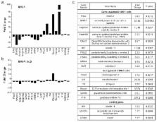
          The laminin-binding integrin α3β1 is highly expressed in epidermal keratinocytes where it regulates both cell-autonomous and paracrine functions that promote wound healing and skin tumorigenesis. However, roles for α3β1 in regulating gene expression programs that control the behaviors of immortalized or transformed keratinocytes remain underexplored. In the current study, we used a microarray approach to identify genes that are regulated by α3β1 in immortalized keratinocytes. α3β1-responsive genes included several that are involved in extracellular matrix proteolysis or remodeling, including fibulin-2 and SPARC. However, α3β1-dependent induction of specific target genes was influenced by the genetic lesion that triggered immortalization, as α3β1-dependent fibulin-2 expression occurred in cells immortalized by either SV40 large T antigen or p53-null mutation, while α3β1-dependent SPARC expression occurred only in the former cells. Interestingly, qPCR arrays did not reveal strong patterns of α3β1-dependent gene expression in freshly isolated primary keratinocytes, suggesting that this regulation is acquired during immortalization. p53-null keratinocytes transformed with oncogenic RasV12 retained α3β1-dependent fibulin-2 expression, and RNAi-mediated knockdown of fibulin-2 in these cells reduced invasion, although not their tumorigenic potential. These findings demonstrate a prominent role for α3β1 in immortalized/transformed keratinocytes in regulating fibulin-2 and other genes that promote matrix remodeling and invasion.

          Related collections

          Most cited references54

          • Record: found
          • Abstract: found
          • Article: not found

          Cancer as an overhealing wound: an old hypothesis revisited.

          What is the relationship between the wound-healing process and the development of cancer? Malignant tumours often develop at sites of chronic injury, and tissue injury has an important role in the pathogenesis of malignant disease, with chronic inflammation being the most important risk factor. The development and functional characterization of genetically modified mice that lack or overexpress genes that are involved in repair, combined with gene-expression analysis in wounds and tumours, have highlighted remarkable similarities between wound repair and cancer. However, a few crucial differences were also observed, which could account for the altered metabolism, impaired differentiation capacity and invasive growth of malignant tumours.
            Bookmark
            • Record: found
            • Abstract: found
            • Article: not found

            Epithelial stem cells, wound healing and cancer.

            It is well established that tissue repair depends on stem cells and that chronic wounds predispose to tumour formation. However, the association between stem cells, wound healing and cancer is poorly understood. Lineage tracing has now shown how stem cells are mobilized to repair skin wounds and how they contribute to skin tumour development. The signalling pathways, including WNT and Hedgehog, that control stem cell behaviour during wound healing are also implicated in tumour formation. Furthermore, tumorigenesis and wound repair both depend on communication between epithelial cells, mesenchymal cells and bone marrow-derived cells. These studies suggest ways to harness stem cells for wound repair while minimizing cancer risk.
              Bookmark
              • Record: found
              • Abstract: found
              • Article: not found

              Automated analysis of retinal images for detection of referable diabetic retinopathy.

              The diagnostic accuracy of computer detection programs has been reported to be comparable to that of specialists and expert readers, but no computer detection programs have been validated in an independent cohort using an internationally recognized diabetic retinopathy (DR) standard. To determine the sensitivity and specificity of the Iowa Detection Program (IDP) to detect referable diabetic retinopathy (RDR). In primary care DR clinics in France, from January 1, 2005, through December 31, 2010, patients were photographed consecutively, and retinal color images were graded for retinopathy severity according to the International Clinical Diabetic Retinopathy scale and macular edema by 3 masked independent retinal specialists and regraded with adjudication until consensus. The IDP analyzed the same images at a predetermined and fixed set point. We defined RDR as more than mild nonproliferative retinopathy and/or macular edema. A total of 874 people with diabetes at risk for DR. Sensitivity and specificity of the IDP to detect RDR, area under the receiver operating characteristic curve, sensitivity and specificity of the retinal specialists' readings, and mean interobserver difference (κ). The RDR prevalence was 21.7% (95% CI, 19.0%-24.5%). The IDP sensitivity was 96.8% (95% CI, 94.4%-99.3%) and specificity was 59.4% (95% CI, 55.7%-63.0%), corresponding to 6 of 874 false-negative results (none met treatment criteria). The area under the receiver operating characteristic curve was 0.937 (95% CI, 0.916-0.959). Before adjudication and consensus, the sensitivity/specificity of the retinal specialists were 0.80/0.98, 0.71/1.00, and 0.91/0.95, and the mean intergrader κ was 0.822. The IDP has high sensitivity and specificity to detect RDR. Computer analysis of retinal photographs for DR and automated detection of RDR can be implemented safely into the DR screening pipeline, potentially improving access to screening and health care productivity and reducing visual loss through early treatment.
                Bookmark

                Author and article information

                Journal
                0426720
                4839
                J Invest Dermatol
                J. Invest. Dermatol.
                The Journal of investigative dermatology
                0022-202X
                1523-1747
                7 May 2014
                02 April 2014
                September 2014
                01 March 2015
                : 134
                : 9
                : 2418-2427
                Affiliations
                [1 ]Center for Cell Biology and Cancer Research, Albany Medical College, MC-165, 47 New Scotland Avenue, Albany, NY, 12208, USA
                [2 ]Center for Functional Genomics, University at Albany, One Discovery Drive, CRC342G, Rensselaer, NY 12144
                Author notes
                []Author for correspondence: C. Michael DiPersio, Ph.D. Current Address: Center for Cell Biology & Cancer Research, Albany Medical College, Mail Code 165, Room MS-420, 47 New Scotland Avenue, Albany, NY 12208-3479, dipersm@ 123456mail.amc.edu , phone: (518) 262-5916, fax: (518) 262-5669
                Article
                NIHMS580381
                10.1038/jid.2014.166
                4134363
                24694902
                86444b08-1c19-4579-87c8-8ddbc5192013
                History
                Categories
                Article

                Dermatology
                α3β1 integrin,keratinocyte,immortalization,epidermis,fibulin-2
                Dermatology
                α3β1 integrin, keratinocyte, immortalization, epidermis, fibulin-2

                Comments

                Comment on this article