70
views
0
recommends
+1 Recommend
0 collections
    0
    shares
      • Record: found
      • Abstract: found
      • Article: not found

      Structures of Hepatitis B Virus Cores Presenting a Model Epitope and Their Complexes with Antibodies

      research-article

      Read this article at
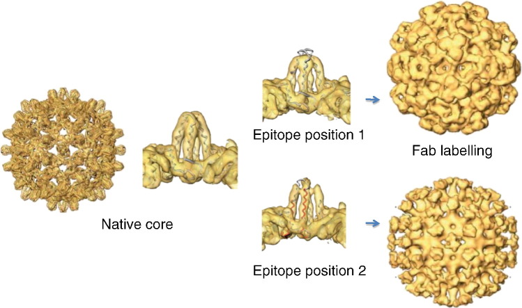
      ScienceOpenPublisherPMC
      Bookmark
          There is no author summary for this article yet. Authors can add summaries to their articles on ScienceOpen to make them more accessible to a non-specialist audience.

          Abstract

          The core shell of hepatitis B virus is a potent immune stimulator, giving a strong neutralizing immune response to foreign epitopes inserted at the immunodominant region, located at the tips of spikes on the exterior of the shell. Here, we analyze structures of core shells with a model epitope inserted at two alternative positions in the immunodominant region. Recombinantly expressed core protein assembles into T = 3 and T = 4 icosahedral shells, and atomic coordinates are available for the T = 4 shell. Since the modified protein assembles predominantly into T = 3 shells, a quasi-atomic model of the native T = 3 shell was made. The spikes in this T = 3 structure resemble those in T = 4 shells crystallized from expressed protein. However, the spikes in the modified shells exhibit an altered conformation, similar to the DNA containing shells in virions. Both constructs allow full access of antibodies to the foreign epitope, DPAFR from the preS1 region of hepatitis B virus surface antigen. However, one induces a 10-fold weaker immune response when injected into mice. In this construct, the epitope is less constrained by the flanking linker regions and is positioned so that the symmetry of the shell causes pairs of epitopes to come close enough to interfere with one another. In the other construct, the epitope mimics the native epitope conformation and position. The interaction of native core shells with an antibody specific to the immunodominant epitope is compared to the constructs with an antibody against the foreign epitope. Our findings have implications for the design of vaccines based on virus-like particles.

          Abstract

          Graphical Abstract

          Highlights

          ► The HBV core shell is highly immunogenic and is being used as a vaccine carrier. ► Insertion of model epitopes into the immunodominant region changes the structure. ► Alternative positions of an epitope give different structures and immunogenicity. ► The structural differences lead to different labeling with antibody fragments. ► We conclude that three‐dimensional structural analysis will be important in vaccine design.

          Related collections

          Most cited references44

          • Record: found
          • Abstract: found
          • Article: not found

          The crystal structure of the human hepatitis B virus capsid.

          Hepatitis B is a small enveloped DNA virus that poses a major hazard to human health. The crystal structure of the T = 4 capsid has been solved at 3.3 A resolution, revealing a largely helical protein fold that is unusual for icosahedral viruses. The monomer fold is stabilized by a hydrophobic core that is highly conserved among human viral variants. Association of two amphipathic alpha-helical hairpins results in formation of a dimer with a four-helix bundle as the major central feature. The capsid is assembled from dimers via interactions involving a highly conserved region near the C terminus of the truncated protein used for crystallization. The major immunodominant region lies at the tips of the alpha-helical hairpins that form spikes on the capsid surface.
            Bookmark
            • Record: found
            • Abstract: found
            • Article: not found

            Determination of the fold of the core protein of hepatitis B virus by electron cryomicroscopy.

            Hepatitis B virus, a major human pathogen with an estimated 300 million carriers worldwide, can lead to cirrhosis and liver cancer in cases of chronic infection. The virus consists of an inner nucleocapsid or core, surrounded by a lipid envelope containing virally encoded surface proteins. The core protein, when expressed in bacteria, assembles into core shell particles, closely resembling the native core of the virus. Here we use electron cryomicroscopy to solve the structure of the core protein to 7.4 A resolution. Images of about 6,400 individual particles from 34 micrographs at different levels of defocus were combined, imposing icosahedral symmetry. The three-dimensional map reveals the complete fold of the polypeptide chain, which is quite unlike previously solved viral capsid proteins and is largely alpha-helical. The dimer clustering of subunits produces spikes on the surface of the shell, which consist of radial bundles of four long alpha-helices. Our model implies that the sequence corresponding to the immunodominant region of the core protein lies at the tip of the spike and also explains other properties of the core protein.
              Bookmark
              • Record: found
              • Abstract: found
              • Article: not found

              MRC image processing programs.

              Digital image processing is an essential step in the determination of macromolecular structures by electron microscopy. Centrally important procedures are the averaging of many images of the subunit to improve the signal, the correction for various transfer functions, and the generation of a three-dimensional map from a set of two-dimensional projections. The detailed way in which these computational procedures are best carried out depends on the symmetry of the object and the type of specimen preparation. Over many years a large set of programs has been written by various members of the Laboratory of Molecular Biology for processing images of two-dimensional crystals and of particles with helical or icosahedral symmetry. The philosophy has been to write stand-alone programs and the whole system is given coherence by the adoption of standard formats for the storage and interchange of different kinds of data. This paper describes the current state of the programs.
                Bookmark

                Author and article information

                Journal
                J Mol Biol
                J. Mol. Biol
                Journal of Molecular Biology
                Elsevier
                0022-2836
                1089-8638
                12 October 2012
                12 October 2012
                : 423
                : 1
                : 63-78
                Affiliations
                [1 ]MRC Laboratory of Molecular Biology, Hills Road, Cambridge CB2 0QH, UK
                [2 ]Faculty of Life Sciences, University of Manchester, Manchester M13 9PT, UK
                [3 ]Latvian Biomedical Research and Study Centre, 1 Ratsupites Street, LV-1067 Riga, Latvia
                Author notes
                [* ]Corresponding author. Faculty of Life Sciences, University of Manchester, Manchester M13 9PT, UK. Alan.Roseman@ 123456manchester.ac.uk
                [1]

                Present addresses: O. Borschukova, 30 Gardner Road, Apt. 3H, Brookline, MA 02445, USA; J. A. Berriman, 51 Oakhill Road, Putney, London SW15 2QJ, UK.

                Article
                YJMBI63735
                10.1016/j.jmb.2012.06.032
                3465560
                22750730
                87d0c9d8-64ba-4ff6-8ed0-2bb7032a5297
                © 2012 Elsevier Ltd.

                This document may be redistributed and reused, subject to certain conditions.

                History
                : 24 February 2012
                : 16 May 2012
                : 20 June 2012
                Categories
                Article

                Molecular biology
                mir, major immunodominant region,em, electron microscopy,fv, fragment of antibody variable region,virus-like particles,image processing,l2, linker 2,3d, three‐dimensional,fab, fragment of antibody,l1, linker 1,cryomicroscopy,vaccine carriers,improved immunogenicity,pbs, phosphate-buffered saline,hbv, hepatitis b virus

                Comments

                Comment on this article