3
views
0
recommends
+1 Recommend
0 collections
    0
    shares
      • Record: found
      • Abstract: found
      • Article: found
      Is Open Access

      Pneumomediastinum and pneumopericardium 11 days after Whipple procedure. A case report and review if the literature

      case-report

      Read this article at

      Bookmark
          There is no author summary for this article yet. Authors can add summaries to their articles on ScienceOpen to make them more accessible to a non-specialist audience.

          Highlights

          • Spontaneous Pneumomediastinum is a rare disease.

          • Combination with Pneumopericardium has not been reported yet.

          • There are no clear guidelines for diagnosis and treatment.

          • Conservative treatment with close monitoring is most of the times sufficient.

          • In surgical patients an upgraded diagnostic and treatment plan could be necessary.

          Abstract

          Introduction

          Spontaneous pneumomediastinum (SPM) is a rare condition classified as free air in the mediastinum in the absence of any precipitating cause. This is the first time that a synchronous presence of pneumopericardium is described. To date, there are no clear guidelines for diagnosis and treatment.

          Presentation of a case

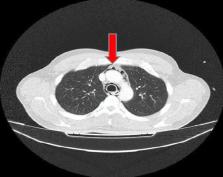
          A 34-year old Caucasian male patient presented to our institution with a recently diagnosed pancreatic adenocarcinoma. He underwent an uneventful pancreaticoduodenectomy (Whipple procedure). Preoperative and intraoperative chest X-Ray after a central line placement were normal. The postoperative course was uneventful, but few hours before his discharge he presented an acute tachycardia and tachypnea with hypocapnia and a transient loss of consciousness. The full-body CT scan revealed a pneumomediastinum and pneumopericardium without any findings of anastomotic leak or other pathology from the abdomen. A meticulous review of the literature was conducted about the pathophysiology, treatment options and outcomes of pneumomediastinum after a surgical procedure.

          Discussion

          This is the first study presenting the case of spontaneous pneumomediastinum with a synchronous pneumopericardium in the literature as a late complication of Whipple procedure. The applied diagnostic algorithm and conservative treatment are presented to extend our limited knowledge about this rare medical entity.

          Conclusion

          Awareness of these medical entities is important for the adequate management and optimal outcome of patients presenting a spontaneous pneumomediastinum. As such, all cases, treatment decisions and outcomes should be reported.

          Related collections

          Most cited references10

          • Record: found
          • Abstract: found
          • Article: not found

          Spontaneous pneumomediastinum: a comparative study and review of the literature.

          Spontaneous pneumomediastinum (SPM) is an unusual occurrence with few cases reported. It is seen after intrathoracic pressure changes leading to alveolar rupture and dissection of air along the tracheobronchial tree. This study was undertaken to provide a thorough clinical and radiologic analysis of this patient population. A retrospective comparative analysis was performed on patients with SPM over 12 years. Patient demographics, clinical presentation, and radiographic and diagnostic studies were recorded. A clinical and radiologic comparison was performed with secondary pneumomediastinum. Seventy-four patients were identified with a diagnosis of pneumomediastinum. A total of 28 patients with SPM were identified. The major initial complaints were chest pain (54%), shortness of breath (39%), and subcutaneous emphysema (32%). The main triggering events were emesis (36%) and asthma flare-ups (21%). No apparent triggering event was noted in 21% of patients. Chest radiograph was diagnostic in 69%; computed tomography was required in 31%. Esophagram, esophagoscopy, and bronchoscopy were performed on an individual basis and were invariably negative. When compared with secondary pneumomediastinum, SPM is more likely to be discovered by chest radiography, has a lower incidence of pneumothorax and pleural effusion, requires a shorter hospital stay, and has no associated mortality. Spontaneous pneumomediastinum is a benign condition that often presents with chest pain or dyspnea. It can develop without a triggering event and with no findings on chest radiography. Treatment is expectant and recurrence is low. Secondary causes must be ruled out to avoid an unfavorable outcome.
            Bookmark
            • Record: found
            • Abstract: not found
            • Article: not found

            Spontaneous mediastinal emphysema

              Bookmark
              • Record: found
              • Abstract: found
              • Article: not found

              Management of spontaneous pneumomediastinum based on clinical experience of 25 cases.

              Spontaneous pneumomediastinum (SPM) is a rare disorder that usually occurs in young individuals and has a self-limiting course and no recurrence in most cases. But actually in many cases, patients are undergo some examinations or some limitations. The purpose of this study was to evaluate the clinical characteristics and recommend appropriate management of SPM. Retrospective research of clinical records of a single institution. Over 11 years, we diagnosed 25 patients (18 males) with SPM. Their average age was 20.1 years (range 13-28 years). Chest pain or neck symptoms were most frequent, and 17 patients (68%) had subcutaneous emphysema. In all cases, blood counts and C-reactive protein (CRP) were measured, and their mean values were 10,100+/-4600/mm(3) and 1.0+/-1.5 mg/dL, respectively. In 20 patients (80%), either leucocytosis or elevated CRP was observed. Twenty-four patients (96%) were admitted (average 7.8+/-4.1 days) and 20 of them were prescribed antibiotics or limited oral intake. The symptoms were self-limiting in all cases and disappeared on average 1.8+/-0.9 days after diagnosis. No recurrence was noted in the approximately 2 years follow-up period. SPM is a self-limiting disease with mild inflammatory signs. For patients suspicious of SPM, shortened hospitalization for about 2 days with observation alone may be feasible, if their symptoms improve gradually. Otherwise, less invasive procedures, such as esophagram, should be performed immediately. Long-term follow-up is usually unnecessary. We propose a new algorithm for management of SPM based on clinical experience.
                Bookmark

                Author and article information

                Contributors
                Journal
                Int J Surg Case Rep
                Int J Surg Case Rep
                International Journal of Surgery Case Reports
                Elsevier
                2210-2612
                08 May 2020
                2020
                08 May 2020
                : 71
                : 27-29
                Affiliations
                [0005]1st Propaedeutic Surgical Department, Hippocratio General Hospital, Medical School, National and Kapodistrian University of Athens, Greece
                Author notes
                [* ]Corresponding author at: 1st Propaedeutic Surgical Clinic, Hippocratio Athens General Hospital, 114 Vassilissis Sophias Avenue, 115 27 Athens, Greece. g.z.vrakopoulou@ 123456gmail.com
                Article
                S2210-2612(20)30204-2
                10.1016/j.ijscr.2020.04.012
                7235923
                32428828
                887e3140-87d4-4768-8168-2b2be8790cd1
                © 2020 The Author(s)

                This is an open access article under the CC BY license (http://creativecommons.org/licenses/by/4.0/).

                History
                : 23 January 2020
                : 30 March 2020
                : 16 April 2020
                Categories
                Article

                spontaneous pneumomediastinum,spontaneous pneumopericardium,postoperative pneumopericardium,pneumomediastinum after whipple,late complication after whipple

                Comments

                Comment on this article