38
views
0
recommends
+1 Recommend
0 collections
    0
    shares
      • Record: found
      • Abstract: found
      • Article: found
      Is Open Access

      Three-dimensional and thermal surface imaging produces reliable measures of joint shape and temperature: a potential tool for quantifying arthritis

      research-article

      Read this article at

      Bookmark
          There is no author summary for this article yet. Authors can add summaries to their articles on ScienceOpen to make them more accessible to a non-specialist audience.

          Abstract

          Introduction

          The assessment of joints with active arthritis is a core component of widely used outcome measures. However, substantial variability exists within and across examiners in assessment of these active joint counts. Swelling and temperature changes, two qualities estimated during active joint counts, are amenable to quantification using noncontact digital imaging technologies. We sought to explore the ability of three dimensional (3D) and thermal imaging to reliably measure joint shape and temperature.

          Methods

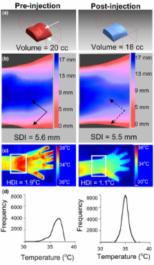
          A Minolta 910 Vivid non-contact 3D laser scanner and a Meditherm med2000 Pro Infrared camera were used to create digital representations of wrist and metacarpalphalangeal (MCP) joints. Specialized software generated 3 quantitative measures for each joint region: 1) Volume; 2) Surface Distribution Index (SDI), a marker of joint shape representing the standard deviation of vertical distances from points on the skin surface to a fixed reference plane; 3) Heat Distribution Index (HDI), representing the standard error of temperatures. Seven wrists and 6 MCP regions from 5 subjects with arthritis were used to develop and validate 3D image acquisition and processing techniques. HDI values from 18 wrist and 9 MCP regions were obtained from 17 patients with active arthritis and compared to data from 10 wrist and MCP regions from 5 controls. Standard deviation (SD), coefficient of variation (CV), and intraclass correlation coefficients (ICC) were calculated for each quantitative measure to establish their reliability. CVs for volume and SDI were <1.3% and ICCs were greater than 0.99.

          Results

          Thermal measures were less reliable than 3D measures. However, significant differences were observed between control and arthritis HDI values. Two case studies of arthritic joints demonstrated quantifiable changes in swelling and temperature corresponding with changes in symptoms and physical exam findings.

          Conclusion

          3D and thermal imaging provide reliable measures of joint volume, shape, and thermal patterns. Further refinement may lead to the use of these technologies to improve the assessment of disease activity in arthritis.

          Related collections

          Most cited references38

          • Record: found
          • Abstract: found
          • Article: not found

          The meaning and use of the area under a receiver operating characteristic (ROC) curve.

          A representation and interpretation of the area under a receiver operating characteristic (ROC) curve obtained by the "rating" method, or by mathematical predictions based on patient characteristics, is presented. It is shown that in such a setting the area represents the probability that a randomly chosen diseased subject is (correctly) rated or ranked with greater suspicion than a randomly chosen non-diseased subject. Moreover, this probability of a correct ranking is the same quantity that is estimated by the already well-studied nonparametric Wilcoxon statistic. These two relationships are exploited to (a) provide rapid closed-form expressions for the approximate magnitude of the sampling variability, i.e., standard error that one uses to accompany the area under a smoothed ROC curve, (b) guide in determining the size of the sample required to provide a sufficiently reliable estimate of this area, and (c) determine how large sample sizes should be to ensure that one can statistically detect differences in the accuracy of diagnostic techniques.
            Bookmark
            • Record: found
            • Abstract: found
            • Article: not found

            The American Rheumatism Association 1987 revised criteria for the classification of rheumatoid arthritis.

            The revised criteria for the classification of rheumatoid arthritis (RA) were formulated from a computerized analysis of 262 contemporary, consecutively studied patients with RA and 262 control subjects with rheumatic diseases other than RA (non-RA). The new criteria are as follows: 1) morning stiffness in and around joints lasting at least 1 hour before maximal improvement; 2) soft tissue swelling (arthritis) of 3 or more joint areas observed by a physician; 3) swelling (arthritis) of the proximal interphalangeal, metacarpophalangeal, or wrist joints; 4) symmetric swelling (arthritis); 5) rheumatoid nodules; 6) the presence of rheumatoid factor; and 7) radiographic erosions and/or periarticular osteopenia in hand and/or wrist joints. Criteria 1 through 4 must have been present for at least 6 weeks. Rheumatoid arthritis is defined by the presence of 4 or more criteria, and no further qualifications (classic, definite, or probable) or list of exclusions are required. In addition, a "classification tree" schema is presented which performs equally as well as the traditional (4 of 7) format. The new criteria demonstrated 91-94% sensitivity and 89% specificity for RA when compared with non-RA rheumatic disease control subjects.
              Bookmark
              • Record: found
              • Abstract: found
              • Article: not found

              American College of Rheumatology. Preliminary definition of improvement in rheumatoid arthritis.

              Trials of rheumatoid arthritis (RA) treatments report the average response in multiple outcome measures for treated patients. It is more clinically relevant to test whether individual patients improve with treatment, and this identifies a single primary efficacy measure. Multiple definitions of improvement are currently in use in different trials. The goal of this study was to promulgate a single definition for use in RA trials. Using the American College of Rheumatology (ACR) core set of outcome measures for RA trials, we tested 40 different definitions of improvement, using a 3-step process. First, we performed a survey of rheumatologists, using actual patient cases from trials, to evaluate which definitions corresponded best to rheumatologists' impressions of improvement, eliminating most candidate definitions of improvement. Second, we tested 20 remaining definitions to determine which maximally discriminated effective treatment from placebo treatment and also minimized placebo response rates. With 8 candidate definitions of improvement remaining, we tested to see which were easiest to use and were best in accord with rheumatologists' impressions of improvement. The following definition of improvement was selected: 20% improvement in tender and swollen joint counts and 20% improvement in 3 of the 5 remaining ACR core set measures: patient and physician global assessments, pain, disability, and an acute-phase reactant. Additional validation of this definition was carried out in a comparative trial, and the results suggest that the definition is statistically powerful and does not identify a large percentage of placebo-treated patients as being improved. We present a definition of improvement which we hope will be used widely in RA trials.
                Bookmark

                Author and article information

                Journal
                Arthritis Res Ther
                Arthritis Research & Therapy
                BioMed Central
                1478-6354
                1478-6362
                2008
                23 January 2008
                : 10
                : 1
                : R10
                Affiliations
                [1 ]Division of Rheumatology, Children's Hospital of Pittsburgh, 3705 Fifth Avenue, University of Pittsburgh School of Medicine, Pittsburgh, PA 15213, USA
                [2 ]Department of Medicine, University of Pittsburgh School of Medicine, 3550 Terrace Street, Pittsburgh, PA 15213, USA
                [3 ]Robotics Institute, Carnegie Mellon University, 5000 Forbes Avenue, Pittsburgh, PA 15213, USA
                Article
                ar2360
                10.1186/ar2360
                2374475
                18215307
                889a3df1-bd10-4dfe-acd7-7addd5ddf1ad
                Copyright © 2008 Spalding et al.; licensee BioMed Central Ltd.

                This is an open access article distributed under the terms of the Creative Commons Attribution License ( http://creativecommons.org/licenses/by/2.0), which permits unrestricted use, distribution, and reproduction in any medium, provided the original work is properly cited.

                History
                : 27 March 2007
                : 8 June 2007
                : 20 June 2007
                : 23 January 2008
                Categories
                Research Article

                Orthopedics
                Orthopedics

                Comments

                Comment on this article