35
views
0
recommends
+1 Recommend
0 collections
    0
    shares
      • Record: found
      • Abstract: found
      • Article: found
      Is Open Access

      Leucocyte-Rich Platelet-Rich Plasma Enhances Fibroblast and Extracellular Matrix Activity: Implications in Wound Healing

      research-article

      Read this article at

      Bookmark
          There is no author summary for this article yet. Authors can add summaries to their articles on ScienceOpen to make them more accessible to a non-specialist audience.

          Abstract

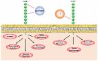
          Background: Platelet-rich plasma (PRP) is an autologous blood product that contains a high concentration of platelets and leucocytes, which are fundamental fibroblast proliferation agents. Literature has emerged that offers contradictory findings about leucocytes within PRP. Herein, we elucidated the effects of highly concentrated leucocytes and platelets on human fibroblasts. Methods: Leucocyte-rich, PRP (LR-PRP) and leucocyte-poor, platelet-poor plasma (LP-PPP) were compared to identify their effects on human fibroblasts, including cell proliferation, wound healing and extracellular matrix and adhesion molecule gene expressions. Results: The LR-PRP exhibited 1422.00 ± 317.21 × 10 3 platelets/µL and 16.36 ± 2.08 × 10 3 white blood cells/µL whilst the LP-PPP demonstrated lower concentrations of 55.33 ± 10.13 × 10 3 platelets/µL and 0.8 ± 0.02 × 10 3 white blood cells/µL. LR-PRP enhanced fibroblast cell proliferation and cell migration, and demonstrated either upregulation or down-regulation gene expression profile of the extracellular matrix and adhesion molecules. Conclusion: LR-PRP has a continuous stimulatory anabolic and ergogenic effect on human fibroblast cells.

          Related collections

          Most cited references98

          • Record: found
          • Abstract: found
          • Article: found
          Is Open Access

          The M1 and M2 paradigm of macrophage activation: time for reassessment

          Macrophages are endowed with a variety of receptors for lineage-determining growth factors, T helper (Th) cell cytokines, and B cell, host, and microbial products. In tissues, macrophages mature and are activated in a dynamic response to combinations of these stimuli to acquire specialized functional phenotypes. As for the lymphocyte system, a dichotomy has been proposed for macrophage activation: classic vs. alternative, also M1 and M2, respectively. In view of recent research about macrophage functions and the increasing number of immune-relevant ligands, a revision of the model is needed. Here, we assess how cytokines and pathogen signals influence their functional phenotypes and the evidence for M1 and M2 functions and revisit a paradigm initially based on the role of a restricted set of selected ligands in the immune response.
            Bookmark
            • Record: found
            • Abstract: found
            • Article: not found

            Classification of platelet concentrates: from pure platelet-rich plasma (P-PRP) to leucocyte- and platelet-rich fibrin (L-PRF).

            The topical use of platelet concentrates is recent and its efficiency remains controversial. Several techniques for platelet concentrates are available; however, their applications have been confusing because each method leads to a different product with different biology and potential uses. Here, we present classification of the different platelet concentrates into four categories, depending on their leucocyte and fibrin content: pure platelet-rich plasma (P-PRP), such as cell separator PRP, Vivostat PRF or Anitua's PRGF; leucocyte- and platelet-rich plasma (L-PRP), such as Curasan, Regen, Plateltex, SmartPReP, PCCS, Magellan or GPS PRP; pure plaletet-rich fibrin (P-PRF), such as Fibrinet; and leucocyte- and platelet-rich fibrin (L-PRF), such as Choukroun's PRF. This classification should help to elucidate successes and failures that have occurred so far, as well as providing an objective approach for the further development of these techniques.
              Bookmark
              • Record: found
              • Abstract: found
              • Article: found
              Is Open Access

              Emerging Roles of Vascular Cell Adhesion Molecule-1 (VCAM-1) in Immunological Disorders and Cancer

              Tumor necrosis factor alpha (TNFα) is a pro-inflammatory cytokine that triggers the expression of inflammatory molecules, including other cytokines and cell adhesion molecules. TNFα induces the expression of intercellular cell adhesion molecule-1 and vascular cell adhesion molecule-1 (VCAM-1). VCAM-1 was originally identified as a cell adhesion molecule that helps regulate inflammation-associated vascular adhesion and the transendothelial migration of leukocytes, such as macrophages and T cells. Recent evidence suggests that VCAM-1 is closely associated with the progression of various immunological disorders, including rheumatoid arthritis, asthma, transplant rejection, and cancer. This review covers the role and relevance of VCAM-1 in inflammation, and also highlights the emerging potential of VCAM-1 as a novel therapeutic target in immunological disorders and cancer.
                Bookmark

                Author and article information

                Journal
                Int J Mol Sci
                Int J Mol Sci
                ijms
                International Journal of Molecular Sciences
                MDPI
                1422-0067
                06 September 2020
                September 2020
                : 21
                : 18
                : 6519
                Affiliations
                [1 ]College of Health and Biomedicine, Victoria University, Melbourne, VIC 3011, Australia; jim.kiatos@ 123456vu.edu.au
                [2 ]Institute for Health and Sport, Victoria University, Melbourne, VIC 3011, Australia; narges.dargahi@ 123456vu.edu.au (N.D.); sarah.fraser@ 123456vu.edu.au (S.F.); kulmira.nurgali@ 123456vu.edu.au (K.N.)
                Author notes
                [* ]Correspondence: jeannie.devereaux@ 123456live.vu.edu.au (J.D.); vasso.apostolopoulos@ 123456vu.edu.au (V.A.); Tel.: +613-83958218 (J.D.); +613-99192025 (V.A.)
                Author information
                https://orcid.org/0000-0003-3214-2938
                https://orcid.org/0000-0002-2597-6929
                https://orcid.org/0000-0002-4707-3739
                https://orcid.org/0000-0001-6788-2771
                Article
                ijms-21-06519
                10.3390/ijms21186519
                7556022
                32900003
                8ba9cfd4-c552-49c8-907a-2ac25d9e0388
                © 2020 by the authors.

                Licensee MDPI, Basel, Switzerland. This article is an open access article distributed under the terms and conditions of the Creative Commons Attribution (CC BY) license ( http://creativecommons.org/licenses/by/4.0/).

                History
                : 27 July 2020
                : 04 September 2020
                Categories
                Article

                Molecular biology
                platelet-rich plasma,prp,platelet gel,platelet-rich fibrin,fibroblasts,extracellular matrix,leucocytes,wound

                Comments

                Comment on this article