29
views
0
recommends
+1 Recommend
0 collections
    0
    shares
      • Record: found
      • Abstract: found
      • Article: found
      Is Open Access

      Dosimetric impact of point A definition on high-dose-rate brachytherapy for cervical cancer: evaluations on conventional point A and MRI-guided, conformal plans

      research-article

      Read this article at

      Bookmark
          There is no author summary for this article yet. Authors can add summaries to their articles on ScienceOpen to make them more accessible to a non-specialist audience.

          Abstract

          Purpose

          To investigate the dosimetric impact of point A definitions on both conventional point A plans and MRI-guided conformal high-dose-rate (HDR) brachytherapy plans.

          Material and methods

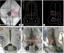
          Fifty-five HDR plans of 36 patients with FIGO stage I to IV cervical cancer were retrospectively studied; these included 30 conventional treatments and 25 conformal plans. Two different point A definitions were explored: the revised Manchester point A and the new point A as recommended by the American Brachytherapy Society. Conventional plans were produced by varying only the point A definition and the normalized isodose lines. Conformal plans were retrospectively generated per GEC-ESTRO recommendations based upon 3.0 Tesla MRI data.

          Results

          Small yet significant variations were found in point A locations (mean: 0.5 cm, maximum: 2.1 cm, p < 0.001). The use of a new point A caused minimal dose variation for both conventional and conformal plans. Conventional plans normalized to the new point A generated up to 12% (avg. 1-3%) higher overall dose in terms of higher total reference air kerma than plans normalized to other points. Dosimetric changes due to point A definitions were up to 11-12% (avg. less than 2%) on target volumes or organs-at-risk.

          Conclusions

          For both conventional and conformal plans, the new point A definition leads to smaller variations caused during implant and/or differences in patient anatomy. Using the new point A is expected to produce more consistent brachytherapy plans and improve outcome analysis.

          Related collections

          Most cited references21

          • Record: found
          • Abstract: found
          • Article: not found

          American Brachytherapy Society consensus guidelines for locally advanced carcinoma of the cervix. Part I: general principles.

          To develop brachytherapy recommendations covering aspects of pretreatment evaluation, treatment, and dosimetric issues for locally advanced cervical cancer. Members of the American Brachytherapy Society (ABS) with expertise in cervical cancer brachytherapy formulated updated recommendations for locally advanced (Federation of Gynecology and Obstetrics Stages IB2-IVA) cervical cancer based on literature review and clinical experience. The ABS recommends the use of brachytherapy as a component of the definitive treatment of locally advanced cervical carcinoma. Precise applicator placement is necessary to maximize the probability of achieving local control without major side effects. The ABS recommends a cumulative delivered dose of approximately 80-90Gy for definitive treatment. The dose delivered to point A should be reported for all brachytherapy applications regardless of treatment-planning technique. The ABS also recommends adoption of the Groupe Européen Curiethérapie-European Society of Therapeutic Radiation Oncology (GEC-ESTRO) guidelines for contouring, image-based treatment planning, and dose reporting. Interstitial brachytherapy may be considered for a small proportion of patients whose disease cannot be adequately encompassed by intracavitary application. It should be performed by practitioners with special expertise in these procedures. Updated ABS recommendations are provided for brachytherapy for locally advanced cervical cancer. Practitioners and cooperative groups are encouraged to use these recommendations to formulate their clinical practices and to adopt dose-reporting policies that are critical for outcome analysis. Copyright © 2012 American Brachytherapy Society. Published by Elsevier Inc. All rights reserved.
            Bookmark
            • Record: found
            • Abstract: found
            • Article: not found

            Computed tomography versus magnetic resonance imaging-based contouring in cervical cancer brachytherapy: results of a prospective trial and preliminary guidelines for standardized contours.

            To compare the contours and dose-volume histograms (DVH) of the tumor and organs at risk (OAR) with computed tomography (CT) vs. magnetic resonance imaging (MRI) in cervical cancer brachytherapy. Ten patients underwent both MRI and CT after applicator insertion. The dose received by at least 90% of the volume (D(90)), the minimal target dose (D(100)), the volume treated to the prescription dose or greater for tumor for the high-risk (HR) and intermediate-risk (IR) clinical target volume (CTV) and the dose to 0.1 cm3, 1 cm3, and 2 cm3 for the OARs were evaluated. A standardized approach to contouring on CT (CT(Std)) was developed, implemented (HR- and IR-CTV(CTStd)), and compared with the MRI contours. Tumor height, thickness, and total volume measurements, as determined by either CT or CT(Std) were not significantly different compared with the MRI volumes. In contrast, the width measurements differed in HR-CTV(CTStd) (p = 0.05) and IR-CTV(CTStd) (p = 0.01). For the HR-CTV(CTStd), this resulted in statistically significant differences in the volume treated to the prescription dose or greater (MRI, 96% vs. CT(Std), 86%, p = 0.01), D(100) (MRI, 5.4 vs. CT(Std), 3.4, p <0.01), and D(90) (MRI, 8.7 vs. CT(Std), 6.7, p <0.01). Correspondingly, the IR-CTV DVH values on MRI vs. CT(Std), differed in the D(100) (MRI, 3.0 vs. CT(Std), 2.2, p = 0.01) and D(90) (MRI, 5.6 vs. CT(Std), 4.6, p = 0.02). The MRI and CT DVH values of the dose to 0.1 cm3, 1 cm3, and 2 cm3 for the OARs were similar. Computed tomography-based or MRI-based scans at brachytherapy are adequate for OAR DVH analysis. However, CT tumor contours can significantly overestimate the tumor width, resulting in significant differences in the D(90), D(100), and volume treated to the prescription dose or greater for the HR-CTV compared with that using MRI. MRI remains the standard for CTV definition.
              Bookmark
              • Record: found
              • Abstract: found
              • Article: not found

              Three-dimensional imaging in gynecologic brachytherapy: a survey of the American Brachytherapy Society.

              To determine current practice patterns with regard to three-dimensional (3D) imaging for gynecologic brachytherapy among American Brachytherapy Society (ABS) members. Registered physician members of the ABS received a 19-item survey by e-mail in August 2007. This report excludes physicians not performing brachytherapy for cervical cancer. Of the 256 surveys sent, we report results for 133 respondents who perform one or more implantations per year for locally advanced cervical cancer. Ultrasound aids 56% of physicians with applicator insertion. After insertion, 70% of physicians routinely obtain a computed tomography (CT) scan. The majority (55%) use CT rather than X-ray films (43%) or magnetic resonance imaging (MRI; 2%) for dose specification to the cervix. However, 76% prescribe to Point A alone instead of using a 3D-derived tumor volume (14%), both Point A and tumor volume (7%), or mg/h (3%). Those using 3D imaging routinely contour the bladder and rectum (94%), sigmoid (45%), small bowel (38%), and/or urethra (8%) and calculate normal tissue dose-volume histogram (DVH) analysis parameters including the D2cc (49%), D1cc (36%), D0.1cc (19%), and/or D5cc (19%). Respondents most commonly modify the treatment plan based on International Commission on Radiation Units bladder and/or rectal point dose values (53%) compared with DVH values (45%) or both (2%). More ABS physician members use CT postimplantation imaging than plain films for visualizing the gynecologic brachytherapy apparatus. However, the majority prescribe to Point A rather than using 3D image based dosimetry. Use of 3D image-based treatment planning for gynecologic brachytherapy has the potential for significant growth in the United States.
                Bookmark

                Author and article information

                Journal
                J Contemp Brachytherapy
                J Contemp Brachytherapy
                JCB
                Journal of Contemporary Brachytherapy
                Termedia Publishing House
                1689-832X
                2081-2841
                28 December 2012
                December 2012
                : 4
                : 4
                : 241-246
                Affiliations
                Radiation Oncology Department, University of Iowa, USA
                Author notes
                Address for correspondence: Yusung Kim, PhD, Ass. Prof., Radiation Oncology Department, University of Iowa, 200 Hawkins Drive, 01607 PFP-W, Iowa City, IOWA 52242-1009, USA. phone: 001 319-384-9406. e-mail: yusung-kim@ 123456uiowa.edu
                Article
                20000
                10.5114/jcb.2012.32559
                3561607
                23378854
                8d5acae7-581a-48bc-a820-07d5cfc5df3c
                Copyright © 2012 Termedia

                This is an Open Access article distributed under the terms of the Creative Commons Attribution-Noncommercial 3.0 Unported License, permitting all non-commercial use, distribution, and reproduction in any medium, provided the original work is properly cited.

                History
                : 19 June 2012
                : 20 October 2012
                : 30 November 2012
                Categories
                Original Paper

                Oncology & Radiotherapy
                brachytherapy,cervical cancer,high-dose-rate brachytherapy,mri-guided,point a

                Comments

                Comment on this article