4
views
0
recommends
+1 Recommend
0 collections
    0
    shares
      • Record: found
      • Abstract: found
      • Article: found
      Is Open Access

      3D-Printing of Hierarchically Designed and Osteoconductive Bone Tissue Engineering Scaffolds

      research-article

      Read this article at

      Bookmark
          There is no author summary for this article yet. Authors can add summaries to their articles on ScienceOpen to make them more accessible to a non-specialist audience.

          Abstract

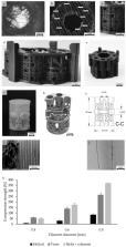
          In Bone Tissue Engineering (BTE), autologous bone-regenerative cells are combined with a scaffold for large bone defect treatment (LBDT). Microporous, polylactic acid (PLA) scaffolds showed good healing results in small animals. However, transfer to large animal models is not easily achieved simply by upscaling the design. Increasing diffusion distances have a negative impact on cell survival and nutrition supply, leading to cell death and ultimately implant failure. Here, a novel scaffold architecture was designed to meet all requirements for an advanced bone substitute. Biofunctional, porous subunits in a load-bearing, compression-resistant frame structure characterize this approach. An open, macro- and microporous internal architecture (100 µm–2 mm pores) optimizes conditions for oxygen and nutrient supply to the implant’s inner areas by diffusion. A prototype was 3D-printed applying Fused Filament Fabrication using PLA. After incubation with Saos-2 (Sarcoma osteogenic) cells for 14 days, cell morphology, cell distribution, cell survival (fluorescence microscopy and LDH-based cytotoxicity assay), metabolic activity (MTT test), and osteogenic gene expression were determined. The adherent cells showed colonization properties, proliferation potential, and osteogenic differentiation. The innovative design, with its porous structure, is a promising matrix for cell settlement and proliferation. The modular design allows easy upscaling and offers a solution for LBDT.

          Related collections

          Most cited references48

          • Record: found
          • Abstract: found
          • Article: not found

          Mechanical properties and the hierarchical structure of bone.

          Detailed descriptions of the structural features of bone abound in the literature; however, the mechanical properties of bone, in particular those at the micro- and nano-structural level, remain poorly understood. This paper surveys the mechanical data that are available, with an emphasis on the relationship between the complex hierarchical structure of bone and its mechanical properties. Attempts to predict the mechanical properties of bone by applying composite rule of mixtures formulae have been only moderately successful, making it clear that an accurate model should include the molecular interactions or physical mechanisms involved in transfer of load across the bone material subunits. Models of this sort cannot be constructed before more information is available about the interactions between the various organic and inorganic components. Therefore, further investigations of mechanical properties at the 'materials level', in addition to the studies at the 'structural level' are needed to fill the gap in our present knowledge and to achieve a complete understanding of the mechanical properties of bone.
            Bookmark
            • Record: found
            • Abstract: found
            • Article: not found

            Scaffolding in tissue engineering: general approaches and tissue-specific considerations.

            B Chan, K Leong (2008)
            Scaffolds represent important components for tissue engineering. However, researchers often encounter an enormous variety of choices when selecting scaffolds for tissue engineering. This paper aims to review the functions of scaffolds and the major scaffolding approaches as important guidelines for selecting scaffolds and discuss the tissue-specific considerations for scaffolding, using intervertebral disc as an example.
              Bookmark
              • Record: found
              • Abstract: found
              • Article: not found

              Fracture healing as a post-natal developmental process: molecular, spatial, and temporal aspects of its regulation.

              Fracture healing is a specialized post-natal repair process that recapitulates aspects of embryological skeletal development. While many of the molecular mechanisms that control cellular differentiation and growth during embryogenesis recur during fracture healing, these processes take place in a post-natal environment that is unique and distinct from those which exist during embryogenesis. This Prospect Article will highlight a number of central biological processes that are believed to be crucial in the embryonic differentiation and growth of skeletal tissues and review the functional role of these processes during fracture healing. Specific aspects of fracture healing that will be considered in relation to embryological development are: (1) the anatomic structure of the fracture callus as it evolves during healing; (2) the origins of stem cells and morphogenetic signals that facilitate the repair process; (3) the role of the biomechanical environment in controlling cellular differentiation during repair; (4) the role of three key groups of soluble factors, pro-inflammatory cytokines, the TGF-beta superfamily, and angiogenic factors, during repair; and (5) the relationship of the genetic components that control bone mass and remodeling to the mechanisms that control skeletal tissue repair in response to fracture. Copyright 2003 Wiley-Liss, Inc.
                Bookmark

                Author and article information

                Journal
                Materials (Basel)
                Materials (Basel)
                materials
                Materials
                MDPI
                1996-1944
                13 April 2020
                April 2020
                : 13
                : 8
                : 1836
                Affiliations
                [1 ]Department of Trauma, Hand and Reconstructive Surgery, University Hospital, Goethe University Frankfurt am Main, 60590 Frankfurt, Germany; Jonas.Neijhoft@ 123456kgu.de (J.N.); Rene.Verboket@ 123456kgu.de (R.D.V.); Johannes.Frank@ 123456kgu.de (J.F.); marzi@ 123456trauma.uni-frankfurt.de (I.M.); d.henrich@ 123456trauma.uni-frankfurt.de (D.H.)
                [2 ]Department of Mechanical Engineering, Institute of Printing Science and Technology, Technical University of Darmstadt, 64289 Darmstadt, Germany; nienhaus@ 123456idd.tu-darmstadt.de (V.N.); valentin.acker@ 123456gmail.com (V.A.); jana.harbig@ 123456googlemail.com (J.H.); fjmenz@ 123456gmail.com (F.M.); jochs-taunus@ 123456gmx.de (J.O.); doersam@ 123456idd.tu-darmstadt.de (E.D.)
                [3 ]Department of Orthopedics and Traumatology, Johannes Gutenberg-University Mainz, 55131 Mainz, Germany; Ulrike.Ritz@ 123456unimedizin-mainz.de
                [4 ]Institute for BioMedical Printing Technology, Technical University of Darmstadt, 64289 Darmstadt, Germany; blaeser@ 123456idd.tu-darmstadt.de
                Author notes
                [* ]Correspondence: Nicolas.Soehling@ 123456kgu.de ; Tel.: +49-69-6301-87373
                [†]

                These authors contributed equally.

                Author information
                https://orcid.org/0000-0003-1086-4016
                https://orcid.org/0000-0001-8936-3227
                https://orcid.org/0000-0002-4338-1777
                https://orcid.org/0000-0002-9728-4032
                Article
                materials-13-01836
                10.3390/ma13081836
                7215341
                32295064
                8db8ddc5-06aa-47cc-a6da-973d8c275214
                © 2020 by the authors.

                Licensee MDPI, Basel, Switzerland. This article is an open access article distributed under the terms and conditions of the Creative Commons Attribution (CC BY) license ( http://creativecommons.org/licenses/by/4.0/).

                History
                : 12 March 2020
                : 08 April 2020
                Categories
                Article

                bone tissue engineering,smart scaffold,scaffold design,osteoconductive

                Comments

                Comment on this article