35
views
0
recommends
+1 Recommend
0 collections
    0
    shares
      • Record: found
      • Abstract: found
      • Article: found
      Is Open Access

      Tissue-specific requirements for specific domains in the FERM protein Moe/Epb4.1l5 during early zebrafish development

      research-article
      1 , 1 ,
      BMC Developmental Biology
      BioMed Central

      Read this article at

      Bookmark
          There is no author summary for this article yet. Authors can add summaries to their articles on ScienceOpen to make them more accessible to a non-specialist audience.

          Abstract

          Background

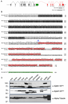
          The FERM domain containing protein Mosaic Eyes (Moe) interacts with Crumbs proteins, which are important regulators of apical identity and size. In zebrafish, loss-of-function mutations in moe result in defects in brain ventricle formation, retinal pigmented epithelium and neural retinal development, pericardial edema, and tail curvature. In humans and mice, there are two major alternately spliced isoforms of the Moe orthologue, Erythrocyte Protein Band 4.1-Like 5 (Epb4.1l5), which we have named Epb4.1l5 long and Epb4.1l5 short, that differ after the FERM domain. Interestingly, Moe and both Epb4.1l5 isoforms have a putative C' terminal Type-I PDZ-Binding Domain (PBD). We previously showed that the N' terminal FERM domain in Moe directly mediates interactions with Crumbs proteins and Nagie oko (Nok) in zebrafish, but the function of the C'terminal half of Moe/Epb4.1l5 has not yet been examined.

          Results

          To define functionally important domains in zebrafish Moe and murine Epb4.1l5, we tested whether injection of mRNAs encoding these proteins could rescue defects in zebrafish moe - embryos. Injection of either moe or epb4.1l5 long mRNA, but not epb4.1l5 short mRNA, could rescue moe - embryonic defects. We also tested whether mRNA encoding C' terminal truncations of Epb4.1l5 long or chimeric constructs with reciprocal swaps of the isoform-specific PBDs could rescue moe - defects. We found that injection of the Epb4.1l5 short chimera (Epb4.1l5 short+long_PBD), containing the PBD from Epb4.1l5 long, could rescue retinal and RPE defects in moe - mutants, but not brain ventricle formation. Injection of the Epb4.1l5 long chimera (Epb4.1l5 long+short_PBD), containing the PBD from Epb4.1l5 short, rescued retinal defects, and to a large extent rescued RPE integrity. The only construct that caused a dominant phenotype in wild-type embryos, was Epb4.1l5 long+short_PBD, which caused brain ventricle defects and edema that were similar to those observed in moe - mutants. Lastly, the morphology of rod photoreceptors in moe - mutants where embryonic defects were rescued by moe or epb4.1l5 long mRNA injection is abnormal and their outer segments are larger than normal.

          Conclusion

          Taken together, the data reveal tissue specificity for the function of the PBD in Epb4.1l5 long, and suggest that additional C' terminal sequences are important for zebrafish retinal development. Additionally, our data provide further evidence that Moe is a negative regulator of rod outer segment size.

          Related collections

          Most cited references32

          • Record: found
          • Abstract: found
          • Article: not found

          crumbs encodes an EGF-like protein expressed on apical membranes of Drosophila epithelial cells and required for organization of epithelia.

          We describe the molecular characterization of the Drosophila gene crumbs, which encodes an integral membrane protein with 30 EGF-like repeats in the extracellular part and exhibits a striking expression pattern. The protein is exclusively localized on the apical membranes of epithelial cells and concentrated at the borders between cells. Mutations in crumbs lead to severe disruptions in the organization of ectodermally derived epithelia and in some cases to cell death in these tissues. The structure and the expression pattern of the protein and the phenotype of mutations indicate a function of crumbs during the development of epithelia, possibly for the establishment and/or maintenance of cell polarity.
            Bookmark
            • Record: found
            • Abstract: found
            • Article: not found

            The development of vision in the zebrafish (Danio rerio).

            We studied the development and maturation of the visual system by determining when zebrafish begin to see and to move their eyes. This information was correlated with the time courses of the development of the retina, the retinofugal projection, the retinal image, and the extraocular muscles, to obtain an integrated picture of early visual development. Two visual behaviors were monitored over 48-96 hr postfertilization (hpf). The startle response (body twitch) was evoked by an abrupt decrease in light intensity. The optokinetic response (tracking eye movements) was evoked by rotation of a striped drum. Visually evoked startle developed over 68-79 hpf, more than 20 hr after the onset of a touch-evoked startle. It was not seen in eyeless fish, excluding a role for nonretinal light senses. Tracking eye movements developed over 73-80 hpf. They were always in the direction of drum rotation, even when the fish had been light deprived from blastula stage, ruling out a "trial and error" period of learning to track the drum. The image formed by the ocular lens was examined in intact fish made transparent by suppressing the formation of melanin. The eye was initially far sighted and gradually improved, so that by 72 hpf the image plane coincided with the photoreceptor layer. The extraocular muscles assumed their adult configuration between 66 and 72 hpf. Thus, the retinal image and functional extraocular muscles appeared nearly simultaneously with the onset of tracking eye movements and probably represent the last events in the construction of this behavior.
              Bookmark
              • Record: found
              • Abstract: found
              • Article: not found

              Mutations in a human homologue of Drosophila crumbs cause retinitis pigmentosa (RP12).

              Retinitis pigmentosa (RP) comprises a clinically and genetically heterogeneous group of diseases that afflicts approximately 1.5 million people worldwide. Affected individuals suffer from a progressive degeneration of the photoreceptors, eventually resulting in severe visual impairment. To isolate candidate genes for chorioretinal diseases, we cloned cDNAs specifically or preferentially expressed in the human retina and the retinal pigment epithelium (RPE) through a novel suppression subtractive hybridization (SSH) method. One of these cDNAs (RET3C11) mapped to chromosome 1q31-q32.1, a region harbouring a gene involved in a severe form of autosomal recessive RP characterized by a typical preservation of the para-arteriolar RPE (RP12; ref. 3). The full-length cDNA encodes an extracellular protein with 19 EGF-like domains, 3 laminin A G-like domains and a C-type lectin domain. This protein is homologous to the Drosophila melanogaster protein crumbs (CRB), and denoted CRB1 (crumbs homologue 1). In ten unrelated RP patients with preserved para-arteriolar RPE, we identified a homozygous AluY insertion disrupting the ORF, five homozygous missense mutations and four compound heterozygous mutations in CRB1. The similarity to CRB suggests a role for CRB1 in cell-cell interaction and possibly in the maintenance of cell polarity in the retina. The distinct RPE abnormalities observed in RP12 patients suggest that CRB1 mutations trigger a novel mechanism of photoreceptor degeneration.
                Bookmark

                Author and article information

                Journal
                BMC Dev Biol
                BMC Developmental Biology
                BioMed Central
                1471-213X
                2008
                11 January 2008
                : 8
                : 3
                Affiliations
                [1 ]Department of Biology and the Molecular and Cellular Biology Program, University of Massachusetts, Amherst, MA, 01003, USA
                Article
                1471-213X-8-3
                10.1186/1471-213X-8-3
                2266719
                18190700
                9154bb5a-097b-4770-94d3-0be86fc0ad6d
                Copyright © 2008 Christensen1 and Jensen; licensee BioMed Central Ltd.

                This is an Open Access article distributed under the terms of the Creative Commons Attribution License ( http://creativecommons.org/licenses/by/2.0), which permits unrestricted use, distribution, and reproduction in any medium, provided the original work is properly cited.

                History
                : 18 September 2007
                : 11 January 2008
                Categories
                Research Article

                Developmental biology
                Developmental biology

                Comments

                Comment on this article