4
views
0
recommends
+1 Recommend
0 collections
    0
    shares
      • Record: found
      • Abstract: found
      • Article: found
      Is Open Access

      Roles and Functions of Exosomal Non-coding RNAs in Vascular Aging

      review-article

      Read this article at

      Bookmark
          There is no author summary for this article yet. Authors can add summaries to their articles on ScienceOpen to make them more accessible to a non-specialist audience.

          Abstract

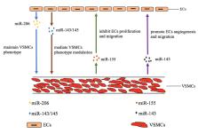
          Aging is a progressive loss of physiological integrity and functionality process which increases susceptibility and mortality to diseases. Vascular aging is a specific type of organic aging. The structure and function changes of endothelial cells (ECs) and vascular smooth muscle cells (VSMCs) are the main cause of vascular aging, which could influence the threshold, process, and severity of vascular related diseases. Accumulating evidences demonstrate that exosomes serve as novel intercellular information communicator between cell to cell by delivering variety biologically active cargos, especially exosomal non-coding RNAs (ncRNAs), which are associated with most of aging-related biological and functional disorders. In this review, we will summerize the emerging roles and mechanisms of exosomal ncRNAs in vascular aging and vascular aging related diseases, focusing on the role of exosomal miRNAs and lncRNAs in regulating the functions of ECs and VSMCs. Moreover, the relationship between the ECs and VSMCs linked by exosomes, the potential diagnostic and therapeutic application of exosomes in vascular aging and the clinical evaluation and treatment of vascular aging and vascular aging related diseases will also be discussed.

          Related collections

          Most cited references110

          • Record: found
          • Abstract: found
          • Article: not found

          General cardiovascular risk profile for use in primary care: the Framingham Heart Study.

          Separate multivariable risk algorithms are commonly used to assess risk of specific atherosclerotic cardiovascular disease (CVD) events, ie, coronary heart disease, cerebrovascular disease, peripheral vascular disease, and heart failure. The present report presents a single multivariable risk function that predicts risk of developing all CVD and of its constituents. We used Cox proportional-hazards regression to evaluate the risk of developing a first CVD event in 8491 Framingham study participants (mean age, 49 years; 4522 women) who attended a routine examination between 30 and 74 years of age and were free of CVD. Sex-specific multivariable risk functions ("general CVD" algorithms) were derived that incorporated age, total and high-density lipoprotein cholesterol, systolic blood pressure, treatment for hypertension, smoking, and diabetes status. We assessed the performance of the general CVD algorithms for predicting individual CVD events (coronary heart disease, stroke, peripheral artery disease, or heart failure). Over 12 years of follow-up, 1174 participants (456 women) developed a first CVD event. All traditional risk factors evaluated predicted CVD risk (multivariable-adjusted P<0.0001). The general CVD algorithm demonstrated good discrimination (C statistic, 0.763 [men] and 0.793 [women]) and calibration. Simple adjustments to the general CVD risk algorithms allowed estimation of the risks of each CVD component. Two simple risk scores are presented, 1 based on all traditional risk factors and the other based on non-laboratory-based predictors. A sex-specific multivariable risk factor algorithm can be conveniently used to assess general CVD risk and risk of individual CVD events (coronary, cerebrovascular, and peripheral arterial disease and heart failure). The estimated absolute CVD event rates can be used to quantify risk and to guide preventive care.
            Bookmark
            • Record: found
            • Abstract: found
            • Article: not found

            MicroRNA-92a controls angiogenesis and functional recovery of ischemic tissues in mice.

            MicroRNAs (miRs) are small noncoding RNAs that regulate gene expression by binding to target messenger RNAs (mRNAs), leading to translational repression or degradation. Here, we show that the miR-17approximately92 cluster is highly expressed in human endothelial cells and that miR-92a, a component of this cluster, controls the growth of new blood vessels (angiogenesis). Forced overexpression of miR-92a in endothelial cells blocked angiogenesis in vitro and in vivo. In mouse models of limb ischemia and myocardial infarction, systemic administration of an antagomir designed to inhibit miR-92a led to enhanced blood vessel growth and functional recovery of damaged tissue. MiR-92a appears to target mRNAs corresponding to several proangiogenic proteins, including the integrin subunit alpha5. Thus, miR-92a may serve as a valuable therapeutic target in the setting of ischemic disease.
              Bookmark
              • Record: found
              • Abstract: found
              • Article: not found

              MicroRNA expression signature and antisense-mediated depletion reveal an essential role of MicroRNA in vascular neointimal lesion formation.

              MicroRNAs (miRNAs) are a recently discovered class of endogenous, small, noncoding RNAs that regulate about 30% of the encoding genes of the human genome. However, the role of miRNAs in vascular disease is currently completely unknown. Using microarray analysis, we demonstrated for the first time that miRNAs are aberrantly expressed in the vascular walls after balloon injury. The aberrantly expressed miRNAs were further confirmed by Northern blot and quantitative real-time polymerase chain reaction. Modulating an aberrantly overexpressed miRNA, miR-21, via antisense-mediated depletion (knock-down) had a significant negative effect on neointimal lesion formation. In vitro, the expression level of miR-21 in dedifferentiated vascular smooth muscle cells was significantly higher than that in fresh isolated differentiated cells. Depletion of miR-21 resulted in decreased cell proliferation and increased cell apoptosis in a dose-dependent manner. MiR-21-mediated cellular effects were further confirmed in vivo in balloon-injured rat carotid arteries. Western blot analysis demonstrated that PTEN and Bcl-2 were involved in miR-21-mediated cellular effects. The results suggest that miRNAs are novel regulatory RNAs for neointimal lesion formation. MiRNAs may be a new therapeutic target for proliferative vascular diseases such as atherosclerosis, postangioplasty restenosis, transplantation arteriopathy, and stroke.
                Bookmark

                Author and article information

                Journal
                Aging Dis
                Aging Dis
                Aging and Disease
                JKL International LLC
                2152-5250
                February 2020
                1 February 2020
                : 11
                : 1
                : 164-178
                Affiliations
                [1-ad-11-1-164]Department of Geriatrics, Institute of Aging and Geriatrics, the Second Xiangya Hospital, Central South University, Changsha, Hunan, 410011, China
                Author notes
                [* ]Correspondence should be addressed to: Dr. You-Shuo Liu. 139 Renmin Middle Road, Changsha, Hunan, 410011, China. E-mail: liuyoushuo@ 123456csu.edu.cn.
                [#]

                These authors contributed equally to this work.

                Article
                ad-11-1-164
                10.14336/AD.2019.0402
                6961769
                32010490
                9404e0e7-eefb-4257-8fde-b59780c5478d
                Copyright: © 2019 Ni et al.

                This is an Open Access article distributed under the terms of the Creative Commons Attribution License, which permits unrestricted use, distribution and reproduction in any medium provided that the original work is properly attributed.

                History
                : 31 January 2019
                : 30 March 2019
                : 2 April 2019
                Categories
                Review Article

                exosomes,endothelial cells,vascular smooth muscle cells,vascular aging,vascular diseases

                Comments

                Comment on this article