34
views
0
recommends
+1 Recommend
0 collections
    0
    shares
      • Record: found
      • Abstract: found
      • Article: not found

      Chlamydia preserves the mitochondrial network necessary for replication via microRNA-dependent inhibition of fission

      research-article

      Read this article at

      ScienceOpenPublisherPMC
      Bookmark
          There is no author summary for this article yet. Authors can add summaries to their articles on ScienceOpen to make them more accessible to a non-specialist audience.

          Abstract

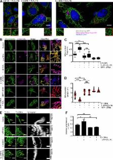
          Chlamydiae are intracellular pathogens that depend on the host for their survival and development. Chowdhury et al. demonstrate that Chlamydia trachomatis infection can prevent mitochondrial fission in primary cells by reducing DRP1 abundance via miR-30c–dependent inhibition of p53.

          Abstract

          Obligate intracellular bacteria such as Chlamydia trachomatis depend on metabolites of the host cell and thus protect their sole replication niche by interfering with the host cells’ stress response. Here, we investigated the involvement of host microRNAs (miRNAs) in maintaining the viability of C. trachomatis–infected primary human cells. We identified miR-30c-5p as a prominently up-regulated miRNA required for the stable down-regulation of p53, a major suppressor of metabolite supply in C. trachomatis–infected cells. Loss of miR-30c-5p led to the up-regulation of Drp1, a mitochondrial fission regulator and a target gene of p53, which, in turn, severely affected chlamydial growth and had a marked effect on the mitochondrial network. Drp1-induced mitochondrial fragmentation prevented replication of C. trachomatis even in p53-deficient cells. Additionally, Chlamydia maintain mitochondrial integrity during reactive oxygen species–induced stress that occurs naturally during infection. We show that C. trachomatis require mitochondrial ATP for normal development and hence postulate that they preserve mitochondrial integrity through a miR-30c-5p–dependent inhibition of Drp1-mediated mitochondrial fission.

          Related collections

          Most cited references68

          • Record: found
          • Abstract: found
          • Article: not found

          Genome sequence of an obligate intracellular pathogen of humans: Chlamydia trachomatis.

          Analysis of the 1,042,519-base pair Chlamydia trachomatis genome revealed unexpected features related to the complex biology of chlamydiae. Although chlamydiae lack many biosynthetic capabilities, they retain functions for performing key steps and interconversions of metabolites obtained from their mammalian host cells. Numerous potential virulence-associated proteins also were characterized. Several eukaryotic chromatin-associated domain proteins were identified, suggesting a eukaryotic-like mechanism for chlamydial nucleoid condensation and decondensation. The phylogenetic mosaic of chlamydial genes, including a large number of genes with phylogenetic origins from eukaryotes, implies a complex evolution for adaptation to obligate intracellular parasitism.
            Bookmark
            • Record: found
            • Abstract: found
            • Article: not found

            Energy substrate modulates mitochondrial structure and oxidative capacity in cancer cells.

            Comparative analysis of cytoplasmic organelles in a variety of tumors relative to normal tissues generally reveals a strong diminution in mitochondrial content and in oxidative phosphorylation capacity. However, little is known about what triggers these modifications and whether or not they are physiologically reversible. We hypothesized that energy substrate availability could play an important role in this phenomenon. The physiological effects of a change in substrate availability were examined on a human cancer cell line (HeLa), focusing specifically on its ability to use glycolysis versus oxidative phosphorylation, and the effect that energy substrate type has on mitochondrial composition, structure, and function. Changes in oxidative phosphorylation were measured in vivo by a variety of techniques, including the use of two novel ratiometric green fluorescent protein biosensors, the expression level of oxidative phosphorylation and some glycolytic enzymes were determined by Western blot, mitochondrial DNA content was measured by real-time PCR, and mitochondrial morphology was monitored by both confocal and electron microscopy. Our data show that the defective mitochondrial system described in cancer cells can be dramatically improved by solely changing substrate availability and that HeLa cells can adapt their mitochondrial network structurally and functionally to derive energy by glutaminolysis only. This could also provide an explanation for the enhancement of oxidative phosphorylation capacity observed after tumor regression or removal. Our work demonstrates that the pleomorphic, highly dynamic structure of the mitochondrion can be remodeled to accommodate a change in oxidative phosphorylation activity. We compared our finding on HeLa cells with those for nontransformed fibroblasts to help distinguish the regulatory pathways.
              Bookmark
              • Record: found
              • Abstract: found
              • Article: found
              Is Open Access

              DIANA miRPath v.2.0: investigating the combinatorial effect of microRNAs in pathways

              MicroRNAs (miRNAs) are key regulators of diverse biological processes and their functional analysis has been deemed central in many research pipelines. The new version of DIANA-miRPath web server was redesigned from the ground-up. The user of DNA Intelligent Analysis (DIANA) DIANA-miRPath v2.0 can now utilize miRNA targets predicted with high accuracy based on DIANA-microT-CDS and/or experimentally verified targets from TarBase v6; combine results with merging and meta-analysis algorithms; perform hierarchical clustering of miRNAs and pathways based on their interaction levels; as well as elaborate sophisticated visualizations, such as dendrograms or miRNA versus pathway heat maps, from an intuitive and easy to use web interface. New modules enable DIANA-miRPath server to provide information regarding pathogenic single nucleotide polymorphisms (SNPs) in miRNA target sites (SNPs module) or to annotate all the predicted and experimentally validated miRNA targets in a selected molecular pathway (Reverse Search module). DIANA-miRPath v2.0 is an efficient and yet easy to use tool that can be incorporated successfully into miRNA-related analysis pipelines. It provides for the first time a series of highly specific tools for miRNA-targeted pathway analysis via a web interface and can be accessed at http://www.microrna.gr/miRPathv2.
                Bookmark

                Author and article information

                Journal
                J Cell Biol
                J. Cell Biol
                jcb
                jcb
                The Journal of Cell Biology
                The Rockefeller University Press
                0021-9525
                1540-8140
                03 April 2017
                : 216
                : 4
                : 1071-1089
                Affiliations
                [1 ]Department of Microbiology, Biocenter, University of Würzburg, 97074 Würzburg, Germany
                [2 ]Institute for Molecular Infection Biology, University of Würzburg, 97080 Würzburg, Germany
                Author notes
                Author information
                http://orcid.org/0000-0002-7355-0674
                http://orcid.org/0000-0001-7051-4670
                http://orcid.org/0000-0002-4581-6244
                http://orcid.org/0000-0003-4740-6991
                Article
                201608063
                10.1083/jcb.201608063
                5379946
                28330939
                96d56153-acd6-45af-9749-b3fb75721756
                © 2017 Chowdhury et al.

                This article is distributed under the terms of an Attribution–Noncommercial–Share Alike–No Mirror Sites license for the first six months after the publication date (see http://www.rupress.org/terms/). After six months it is available under a Creative Commons License (Attribution–Noncommercial–Share Alike 4.0 International license, as described at https://creativecommons.org/licenses/by-nc-sa/4.0/).

                History
                : 19 August 2016
                : 09 December 2016
                : 15 February 2017
                Funding
                Funded by: Deutsche Forschungsgemeinschaft, DOI https://doi.org/10.13039/501100001659;
                Award ID: SPP1580
                Award ID: Infect-ERA CINOCA
                Categories
                Research Articles
                Article
                19
                42

                Cell biology
                Cell biology

                Comments

                Comment on this article