31
views
0
recommends
+1 Recommend
0 collections
    0
    shares
      • Record: found
      • Abstract: found
      • Article: found
      Is Open Access

      Intra-islet endothelial cell and β-cell crosstalk: Implication for islet cell transplantation

      review-article

      Read this article at

      Bookmark
          There is no author summary for this article yet. Authors can add summaries to their articles on ScienceOpen to make them more accessible to a non-specialist audience.

          Abstract

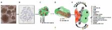
          The intra-islet microvasculature is a critical interface between the blood and islet endocrine cells governing a number of cellular and pathophysiological processes associated with the pancreatic tissue. A growing body of evidence indicates a strong functional and physical interdependency of β-cells with endothelial cells (ECs), the building blocks of islet microvasculature. Intra-islet ECs, actively regulate vascular permeability and appear to play a role in fine-tuning blood glucose sensing and regulation. These cells also tend to behave as “guardians”, controlling the expression and movement of a number of important immune mediators, thereby strongly contributing to the physiology of islets. This review will focus on the molecular signalling and crosstalk between the intra-islet ECs and β-cells and how their relationship can be a potential target for intervention strategies in islet pathology and islet transplantation.

          Related collections

          Most cited references147

          • Record: found
          • Abstract: found
          • Article: not found

          Membrane-derived microvesicles: important and underappreciated mediators of cell-to-cell communication.

          Normal and malignant cells shed from their surface membranes as well as secrete from the endosomal membrane compartment circular membrane fragments called microvesicles (MV). MV that are released from viable cells are usually smaller in size compared to the apoptotic bodies derived from damaged cells and unlike them do not contain fragmented DNA. Growing experimental evidence indicates that MV are an underappreciated component of the cell environment and play an important pleiotropic role in many biological processes. Generally, MV are enriched in various bioactive molecules and may (i) directly stimulate cells as a kind of 'signaling complex', (ii) transfer membrane receptors, proteins, mRNA and organelles (e.g., mitochondria) between cells and finally (iii) deliver infectious agents into cells (e.g., human immuno deficiency virus, prions). In this review, we discuss the pleiotropic effects of MV that are important for communication between cells, as well as the role of MV in carcinogenesis, coagulation, immune responses and modulation of susceptibility/infectability of cells to retroviruses or prions.
            Bookmark
            • Record: found
            • Abstract: found
            • Article: not found

            Angiopoietin-2, a natural antagonist for Tie2 that disrupts in vivo angiogenesis.

            Angiogenesis is thought to depend on a precise balance of positive and negative regulation. Angiopoietin-1 (Ang1) is an angiogenic factor that signals through the endothelial cell-specific Tie2 receptor tyrosine kinase. Like vascular endothelial growth factor, Ang1 is essential for normal vascular development in the mouse. An Ang1 relative, termed angiopoietin-2 (Ang2), was identified by homology screening and shown to be a naturally occurring antagonist for Ang1 and Tie2. Transgenic overexpression of Ang2 disrupts blood vessel formation in the mouse embryo. In adult mice and humans, Ang2 is expressed only at sites of vascular remodeling. Natural antagonists for vertebrate receptor tyrosine kinases are atypical; thus, the discovery of a negative regulator acting on Tie2 emphasizes the need for exquisite regulation of this angiogenic receptor system.
              Bookmark
              • Record: found
              • Abstract: found
              • Article: not found

              Vascular permeability factor/vascular endothelial growth factor: a critical cytokine in tumor angiogenesis and a potential target for diagnosis and therapy.

              Vascular endothelial growth factor A (VEGF-A), the founding member of the vascular permeability factor (VPF)/VEGF family of proteins, is an important angiogenic cytokine with critical roles in tumor angiogenesis. This article reviews the literature with regard to VEGF-A's multiple functions, the mechanisms by which it induces angiogenesis, and its current and projected roles in clinical oncology. VEGF-A is a multifunctional cytokine that is widely expressed by tumor cells and that acts through receptors (VEGFR-1, VEGFR-2, and neuropilin) that are expressed on vascular endothelium and on some other cells. It increases microvascular permeability, induces endothelial cell migration and division, reprograms gene expression, promotes endothelial cell survival, prevents senescence, and induces angiogenesis. Recently, VEGF-A has also been shown to induce lymphangiogenesis. Measurements of circulating levels of VEGF-A may have value in estimating prognosis, and VEGF-A and its receptors are potential targets for therapy. Recognized as the single most important angiogenic cytokine, VEGF-A has a central role in tumor biology and will likely have an important role in future approaches designed to evaluate patient prognosis. It may also become an important target for cancer therapy.
                Bookmark

                Author and article information

                Journal
                World J Transplant
                WJT
                World Journal of Transplantation
                Baishideng Publishing Group Inc
                2220-3230
                24 April 2017
                24 April 2017
                : 7
                : 2
                : 117-128
                Affiliations
                Siddharth Narayanan, Gopalakrishnan Loganathan, Maheswaran Dhanasekaran, William Tucker, Ankit Patel, SriPrakash Mokshagundam, Michael G Hughes, Stuart K Williams, Appakalai N Balamurugan, Clinical Islet Cell Laboratory, Center for Cellular Transplantation, Cardiovascular Innovation Institute, Department of Surgery, University of Louisville, Louisville, KY 40202, United States
                Venugopal Subhashree, School of Biosciences and Technology, VIT University, Vellore 632014, India
                Author notes

                Author contributions: All authors contributed to this paper.

                Correspondence to: Appakalai N Balamurugan, PhD, Clinical Islet Cell Laboratory, Center for Cellular Transplantation, Cardiovascular Innovation Institute, Department of Surgery, University of Louisville, 302 E Muhammad Ali Blvd., Louisville, KY 40202, United States. bala.appakalai@ 123456louisville.edu

                Telephone: +1-502-7947070 Fax: +1-502-8540558

                Article
                jWJT.v7.i2.pg117
                10.5500/wjt.v7.i2.117
                5409911
                28507914
                96fbf391-bc71-47af-80cf-b5fc9ec46d45
                ©The Author(s) 2017. Published by Baishideng Publishing Group Inc. All rights reserved.

                Open-Access: This article is an open-access article which was selected by an in-house editor and fully peer-reviewed by external reviewers. It is distributed in accordance with the Creative Commons Attribution Non Commercial (CC BY-NC 4.0) license, which permits others to distribute, remix, adapt, build upon this work non-commercially, and license their derivative works on different terms, provided the original work is properly cited and the use is non-commercial. See: http://creativecommons.org/licenses/by-nc/4.0/

                History
                : 4 January 2017
                : 28 February 2017
                : 23 March 2017
                Categories
                Review

                islets,endothelial cells,islet cell transplantation,beta-cells,microvasculature,paracrine signalling

                Comments

                Comment on this article