11
views
0
recommends
+1 Recommend
0 collections
    0
    shares
      • Record: found
      • Abstract: found
      • Article: found
      Is Open Access

      ACUTE CENTRAL SEROUS CHORIORETINOPATHY : Factors Influencing Episode Duration

      research-article

      Read this article at

      Bookmark
          There is no author summary for this article yet. Authors can add summaries to their articles on ScienceOpen to make them more accessible to a non-specialist audience.

          Abstract

          The investigation of clinical and multimodal imaging factors influencing the duration of first, acute, and treatment-naive CSCR episodes by survival analysis showed that higher subfoveal choroidal thickness, higher pigment epithelial detachment or bump at leakage sites, and older age were independent predictors of longer episodes.

          Abstract

          Purpose:

          To evaluate the influence of clinical and multimodal imaging parameters on the duration of acute central serous chorioretinopathy (CSCR) episodes.

          Methods:

          Consecutive patients with first, treatment-naïve central serous chorioretinopathy episodes presenting within 20 days of symptoms onset were prospectively included. They were reevaluated 15 days to 20 days later, followed by monthly evaluation for 6 months. Subfoveal choroidal thickness (SFCT), fluorescein leakage intensity on fluorescein angiography, elevation of retinal pigment epithelium (RPE) lesions at leakage sites, focal/multifocal pattern of indocyanine green angiography (ICGA) at baseline, time-dependent pattern of subretinal fluid (SRF) resorption on OCT using volume segmentation, history of corticosteroid intake and mean blood pressure were evaluated using univariate (Log rank test) and multivariate (Cox proportional hazard regression) survival analysis.

          Results:

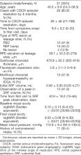
          Thirty-one patients were included (26 men, 5 women, mean age: 40.0 ± 8.9 years, range: 24–58), of which 26 (84%) had episode resolution by 6 months. Using univariate analysis, episode duration was longer in cases with subfoveal choroidal thickness ≥500 μm ( P = 0.0002), retinal pigment epithelium elevation at leakage sites ≥50 μm ( P = 0.033), and a peak in subretinal fluid observed during follow-up ( P = 0.013), and there was a near-significant association of intense fluorescein leakage ( P = 0.074) with longer episodes. Using multivariate analysis, subfoveal choroidal thickness ≥500 μm ( P = 0.017), retinal pigment epithelium elevation at leakage sites ≥50 μm ( P = 0.010) and patient age ≥40 years ( P = 0.010) were significantly and independently associated to longer episodes. Indocyanine green angiography pattern, corticosteroid intake, and blood pressure did not influence episode duration.

          Conclusion:

          Older age, higher subfoveal choroidal thickness, and higher degree of retinal pigment epithelium alteration at leakage sites are independent factors of longer acute central serous chorioretinopathy episodes.

          Related collections

          Most cited references50

          • Record: found
          • Abstract: found
          • Article: not found

          Central serous chorioretinopathy: Recent findings and new physiopathology hypothesis.

          Central serous chorioretinopathy (CSCR) is a major cause of vision threat among middle-aged male individuals. Multimodal imaging led to the description of a wide range of CSCR manifestations, and highlighted the contribution of the choroid and pigment epithelium in CSCR pathogenesis. However, the exact molecular mechanisms of CSCR have remained uncertain. The aim of this review is to recapitulate the clinical understanding of CSCR, with an emphasis on the most recent findings on epidemiology, risk factors, clinical and imaging diagnosis, and treatments options. It also gives an overview of the novel mineralocorticoid pathway hypothesis, from animal data to clinical evidences of the biological efficacy of oral mineralocorticoid antagonists in acute and chronic CSCR patients. In rodents, activation of the mineralocorticoid pathway in ocular cells either by intravitreous injection of its specific ligand, aldosterone, or by over-expression of the receptor specifically in the vascular endothelium, induced ocular phenotypes carrying many features of acute CSCR. Molecular mechanisms include expression of the calcium-dependent potassium channel (KCa2.3) in the endothelium of choroidal vessels, inducing subsequent vasodilation. Inappropriate or over-activation of the mineralocorticoid receptor in ocular cells and other tissues (such as brain, vessels) could link CSCR with the known co-morbidities observed in CSCR patients, including hypertension, coronary disease and psychological stress.
            Bookmark
            • Record: found
            • Abstract: found
            • Article: not found

            EN FACE IMAGING OF PACHYCHOROID SPECTRUM DISORDERS WITH SWEPT-SOURCE OPTICAL COHERENCE TOMOGRAPHY.

            To correlate clinical manifestations with choroidal morphology in pachychoroid disorders, including central serous chorioretinopathy, pachychoroid pigment epitheliopathy, pachychoroid neovasculopathy, and polypoidal choroidal vasculopathy, using en face swept-source optical coherence tomography (OCT).
              Bookmark
              • Record: found
              • Abstract: found
              • Article: not found

              Central serous chorioretinopathy in younger and older adults.

              The purpose of the study is to investigate the demographic characteristics and clinical findings of central serous chorioretinopathy (CSC). This study examined a consecutive series of 130 patients with CSC seen over an 18-month period. The mean age of the patients when examined was 51 years, and the male-to-female ratio was 2.6:1.0. A total of 62 patients were older than 50 years of age when first examined. Although the patients shared some clinical and angiographic similarities, the older patients had a lower mean visual acuity and were more likely to have diffuse retinal pigment epitheliopathy, bilateral involvement, and secondary choroidal neovascularization than were the younger patients. With ophthalmoscopic and angiographic examination results, it was possible to differentiate CSC in older adults from choroidal neovascularization. This study expands the clinical concept of CSC. The male-to-female ratio was much lower, and the range of ages of the patients was much greater than in previous studies. Disease manifestations in older adults differed somewhat from those seen in younger adults. In older patients, CSC can be distinguished from other exudative maculopathies, particularly that of choroidal neovascularization secondary to age-related macular degeneration.
                Bookmark

                Author and article information

                Journal
                Retina
                Retina (Philadelphia, Pa.)
                retina
                Retina (Philadelphia, Pa.)
                Retina
                0275-004X
                1539-2864
                October 2017
                06 October 2017
                : 37
                : 10
                : 1905-1915
                Affiliations
                [* ]Department of Ophthalmology, University of Lausanne, Jules-Gonin Eye Hospital, Fondation Asile des Aveugles, Lausanne, Switzerland;
                []Sorbonne Universités, UPMC Université Paris 06, UMR 1138, Centre de Recherche des Cordeliers, Team 1 and 17, Paris, France;
                []INSERM, UMR 1138, Centre de Recherche des Cordeliers, Paris, France; and
                [§ ]Université Paris Descartes, Sorbonne Paris Cité, UMR 1138, Centre de Recherche des Cordeliers, Paris, France.
                Author notes
                Reprint requests: Francine Behar-Cohen, MD, PhD, Department of Ophthalmology, University of Lausanne, Jules-Gonin Eye Hospital, Fondation Asile des Aveugles Avenue de France 15, 1004 Lausanne, Switzerland; e-mail: francine.behar@ 123456gmail.com
                Article
                Retina-216-0870 00013
                10.1097/IAE.0000000000001443
                5642321
                28067724
                979f1db5-3b6a-4390-8688-fb757a021e88
                Copyright @ 2017

                This is an open-access article distributed under the terms of the Creative Commons Attribution-Non Commercial-No Derivatives License 4.0 (CCBY-NC-ND), where it is permissible to download and share the work provided it is properly cited. The work cannot be changed in any way or used commercially without permission from the journal.

                History
                Categories
                Original Study
                Custom metadata
                TRUE

                central serous chorioretinopathy,choroid,retinal pigment epithelium,choroidal thickness,optical coherence tomography,fluorescein angiography,indocyanine green angiography,age factors,time factors,steroids

                Comments

                Comment on this article