12
views
0
recommends
+1 Recommend
0 collections
    0
    shares
      • Record: found
      • Abstract: found
      • Article: found
      Is Open Access

      Administration of hydrogen-rich water prevents vascular aging of the aorta in LDL receptor-deficient mice

      research-article

      Read this article at

      Bookmark
          There is no author summary for this article yet. Authors can add summaries to their articles on ScienceOpen to make them more accessible to a non-specialist audience.

          Abstract

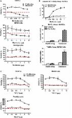
          The main cause of arteriosclerosis is atherosclerosis in the aorta. Atherosclerosis is recognized as a chronic inflammatory condition that begins with the dysfunction or activation of arterial endothelium. Low-density lipoprotein (LDL) and especially its oxidized form play a key role in endothelial dysfunction and atherogenesis. Recent studies showed that senescent cells are involved in the development and progression of atherosclerosis, and eliminating senescent cells suppresses the senescence-associated secretory phenotype. We previously reported that molecular hydrogen-rich water (HW) has antioxidant and anti-inflammatory effects in numerous diseases. Here, we used LDL receptor-deficient mice fed a high-fat diet (HFD) for 13 weeks as a model for atherosclerosis and evaluated the effects of continuous administration of HW. The numbers of endothelial cells in the atheroma expressing the senescence factors p16 INK4a and p21 decreased in HFD-fed mice given HW compared with HFD-fed mice given control water. Furthermore, macrophage infiltration and Tnfα expression in the atheroma were also suppressed. These results suggest that vascular aging can be suppressed by HW.

          Related collections

          Most cited references37

          • Record: found
          • Abstract: found
          • Article: not found

          Cellular senescence in aging and age-related disease: from mechanisms to therapy.

          Cellular senescence, a process that imposes permanent proliferative arrest on cells in response to various stressors, has emerged as a potentially important contributor to aging and age-related disease, and it is an attractive target for therapeutic exploitation. A wealth of information about senescence in cultured cells has been acquired over the past half century; however, senescence in living organisms is poorly understood, largely because of technical limitations relating to the identification and characterization of senescent cells in tissues and organs. Furthermore, newly recognized beneficial signaling functions of senescence suggest that indiscriminately targeting senescent cells or modulating their secretome for anti-aging therapy may have negative consequences. Here we discuss current progress and challenges in understanding the stressors that induce senescence in vivo, the cell types that are prone to senesce, and the autocrine and paracrine properties of senescent cells in the contexts of aging and age-related diseases as well as disease therapy.
            Bookmark
            • Record: found
            • Abstract: found
            • Article: not found

            Persistent DNA damage signaling triggers senescence-associated inflammatory cytokine secretion

            Cellular senescence suppresses cancer by stably arresting the proliferation of damaged cells1. Paradoxically, senescent cells also secrete factors that alter tissue microenvironments2. The pathways regulating this secretion are unknown. We show that damaged human cells develop persistent chromatin lesions bearing hallmarks of DNA double-strand breaks (DSBs), which initiate increased secretion of inflammatory cytokines such as interleukin-6 (IL-6). Cytokine secretion occurred only after establishment of persistent DNA damage signaling, usually associated with senescence, not after transient DNA damage responses (DDR). Initiation and maintenance of this cytokine response required the DDR proteins ATM, NBS1 and CHK2, but not the cell cycle arrest enforcers p53 and pRb. ATM was also essential for IL-6 secretion during oncogene-induced senescence and by damaged cells that bypass senescence. Further, DDR activity and IL-6 were elevated in human cancers, and ATM-depletion suppressed the ability of senescent cells to stimulate IL-6-dependent cancer cell invasiveness. Thus, in addition to orchestrating cell cycle checkpoints and DNA repair, a novel and important role of the DDR is to allow damaged cells to communicate their compromised state to the surrounding tissue.
              Bookmark
              • Record: found
              • Abstract: found
              • Article: found
              Is Open Access

              Identification of a novel senolytic agent, navitoclax, targeting the Bcl‐2 family of anti‐apoptotic factors

              Summary Clearing senescent cells extends healthspan in mice. Using a hypothesis‐driven bioinformatics‐based approach, we recently identified pro‐survival pathways in human senescent cells that contribute to their resistance to apoptosis. This led to identification of dasatinib (D) and quercetin (Q) as senolytics, agents that target some of these pathways and induce apoptosis preferentially in senescent cells. Among other pro‐survival regulators identified was Bcl‐xl. Here, we tested whether the Bcl‐2 family inhibitors, navitoclax (N) and TW‐37 (T), are senolytic. Like D and Q, N is senolytic in some, but not all types of senescent cells: N reduced viability of senescent human umbilical vein epithelial cells (HUVECs), IMR90 human lung fibroblasts, and murine embryonic fibroblasts (MEFs), but not human primary preadipocytes, consistent with our previous finding that Bcl‐xl siRNA is senolytic in HUVECs, but not preadipocytes. In contrast, T had little senolytic activity. N targets Bcl‐2, Bcl‐xl, and Bcl‐w, while T targets Bcl‐2, Bcl‐xl, and Mcl‐1. The combination of Bcl‐2, Bcl‐xl, and Bcl‐w siRNAs was senolytic in HUVECs and IMR90 cells, while combination of Bcl‐2, Bcl‐xl, and Mcl‐1 siRNAs was not. Susceptibility to N correlated with patterns of Bcl‐2 family member proteins in different types of human senescent cells, as has been found in predicting response of cancers to N. Thus, N is senolytic and acts in a potentially predictable cell type‐restricted manner. The hypothesis‐driven, bioinformatics‐based approach we used to discover that dasatinib (D) and quercetin (Q) are senolytic can be extended to increase the repertoire of senolytic drugs, including additional cell type‐specific senolytic agents.
                Bookmark

                Author and article information

                Contributors
                iohsawa@tmig.or.jp
                Journal
                Sci Rep
                Sci Rep
                Scientific Reports
                Nature Publishing Group UK (London )
                2045-2322
                14 November 2018
                14 November 2018
                2018
                : 8
                : 16822
                Affiliations
                [1 ]ISNI 0000 0000 9337 2516, GRID grid.420122.7, Biological Process of Aging, Tokyo Metropolitan Institute of Gerontology, ; Tokyo, Japan
                [2 ]ISNI 0000 0004 1762 8507, GRID grid.265125.7, Department of Life Sciences, Toyo University, ; Gunma, Japan
                [3 ]ISNI 0000 0001 2173 8328, GRID grid.410821.e, Department of Ophthalmology, Nippon Medical School, ; Tokyo, Japan
                [4 ]Central Research Institute, ITO EN Ltd., Makinohara, Japan
                Article
                35239
                10.1038/s41598-018-35239-0
                6235982
                30429524
                97c56ada-b9b4-4cba-9d16-819cba85f0b7
                © The Author(s) 2018

                Open Access This article is licensed under a Creative Commons Attribution 4.0 International License, which permits use, sharing, adaptation, distribution and reproduction in any medium or format, as long as you give appropriate credit to the original author(s) and the source, provide a link to the Creative Commons license, and indicate if changes were made. The images or other third party material in this article are included in the article’s Creative Commons license, unless indicated otherwise in a credit line to the material. If material is not included in the article’s Creative Commons license and your intended use is not permitted by statutory regulation or exceeds the permitted use, you will need to obtain permission directly from the copyright holder. To view a copy of this license, visit http://creativecommons.org/licenses/by/4.0/.

                History
                : 17 July 2018
                : 25 October 2018
                Funding
                Funded by: Research Center for Biomedical Engineering in Toyo University
                Funded by: FundRef https://doi.org/10.13039/501100001691, Japan Society for the Promotion of Science (JSPS);
                Award ID: 16H03267
                Award ID: 18K11092
                Award Recipient :
                Categories
                Article
                Custom metadata
                © The Author(s) 2018

                Uncategorized
                Uncategorized

                Comments

                Comment on this article