18
views
0
recommends
+1 Recommend
0 collections
    0
    shares
      • Record: found
      • Abstract: found
      • Article: found
      Is Open Access

      Cross-Sectional Inverse Associations of Obesity and Fat Accumulation Indicators with Testosterone in Non-Diabetic Aging Men

      research-article

      Read this article at

      Bookmark
          There is no author summary for this article yet. Authors can add summaries to their articles on ScienceOpen to make them more accessible to a non-specialist audience.

          Abstract

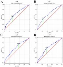
          Introduction/Objective: The aim of the study was to show which of the adipose tissue accumulation indicators correlate with testosterone disorders in non-diabetic aging men. Material and methods: 455 non diabetic men, recruited at primary care facilities, aged 50–75 participated in the study. The participants underwent anthropometric measurement and ELISA determination of total testosterone (TT), estradiol (E 2), dehydroepiandrosterone sulphate (DHEA-S), sex hormone binding protein (SHBG), and the determination of fasting glucose (FPG), high-density lipids cholesterol (HDL-Ch), and triacylglycerols (TAG) in serum. The following indicators were calculated: body mass index (BMI), waist-to-hip ratio (WHR), lipid accumulation product (LAP), and visceral adiposity index (VAI). Results: Men with testosterone deficiency syndrome (TDS) differed in each of the assessed obesity indices from those without TDS. All of the studied parameters correlated significantly negatively with TT concentration in blood serum, with VAI being the strongest predictor of TDS. It was shown that the threshold value at which the risk of TDS increased was 28.41 kg/m 2 for BMI, 1.58 for VAI, 104 cm for WC, and 37.01 for LAP. Conclusions: Indicators of fat accumulation that take into account biochemical parameters in assessing lipid metabolism are better markers of actual body fat deposition than indicators based solely on anthropometric measurements. Among them, VAI seems the most suitable biomarker of TDS in non-diabetic aging men.

          Related collections

          Most cited references24

          • Record: found
          • Abstract: found
          • Article: not found

          Testosterone: a vascular hormone in health and disease.

          Coronary heart disease is a leading cause of premature death in men. Epidemiological studies have shown a high prevalence of low serum testosterone levels in men with cardiovascular disease (CVD). Furthermore, a low testosterone level is associated in some but not in all observational studies with an increase in cardiovascular events and mortality. Testosterone has beneficial effects on several cardiovascular risk factors, which include cholesterol, endothelial dysfunction and inflammation: key mediators of atherosclerosis. A bidirectional relationship between low endogenous testosterone levels and concurrent illness complicates attempts to validate causality in this association and potential mechanistic actions are complex. Testosterone is a vasoactive hormone that predominantly has vasodilatory actions on several vascular beds, although some studies have reported conflicting effects. In clinical studies, acute and chronic testosterone administration increases coronary artery diameter and flow, improves cardiac ischaemia and symptoms in men with chronic stable angina and reduces peripheral vascular resistance in chronic heart failure. Although the mechanism of the action of testosterone on vascular tone in vivo is not understood, laboratory research has found that testosterone is an L-calcium channel blocker and induces potassium channel activation in vascular smooth muscle cells. Animal studies have consistently demonstrated that testosterone is atheroprotective, whereas testosterone deficiency promotes the early stages of atherogenesis. The translational effects of testosterone between in vitro animal and human studies, some of which have conflicting effects, will be discussed in this review. We review the evidence for a role of testosterone in vascular health, its therapeutic potential and safety in hypogonadal men with CVD, and some of the possible underlying mechanisms.
            Bookmark
            • Record: found
            • Abstract: found
            • Article: not found

            The hypogonadal-obesity cycle: role of aromatase in modulating the testosterone-estradiol shunt--a major factor in the genesis of morbid obesity.

            Massive obesity in males is associated with decreased total and free testosterone levels as well as elevated estradiol levels. The decrease in testosterone occurs without the compensatory increases in gonadotropin and a progressive hypogonadotropic hypogonadal cycle develops. During the hypogonadal state, there is a preferential deposition of abdominal adipose tissue. With the increasing fatty-tissue accumulation, there is an increase of aromatase activity that is associated with a greater conversion of testosterone to estradiol (testosterone-estradiol shunt). This results in further depression of testosterone concentrations and leads to the increased preferential deposition of abdominal fat that, in turn, leads to a progressive hypogonadal state. Testalactone, an aromatase inhibitor, interrupts this cycle and repairs the depressed testosterone concentrations and decreases estradiol levels. This increases the testosterone levels and reverses the preferential deposition of abdominal fat, while increasing muscle protein and fat-free mass.
              Bookmark
              • Record: found
              • Abstract: found
              • Article: not found

              Obesity and late-onset hypogonadism.

              Obesity and male hypogonadism (HG) are often associated, as demonstrated in all cross-sectional studies. Prospective studies have indicated that i) having HG at baseline increases the risk of visceral obesity (and metabolic syndrome) and that ii) obesity induces incident HG. Hence, there is a bidirectional relationship between the two conditions. This is the main topic of this review, along with some pathogenic considerations. Meta-analysis of intervention studies indicates that treating obesity is a very efficient treatment for obesity-induced HG. The mechanism by which obesity induces HG has not yet been completely understood, but dietary-induced hypothalamic inflammation, along with a decreased GnRH release, is plausible. Among patients seeking medical care for obesity, the proportion of HG is relatively high. The prevalence of obesity among patients referring for sexual dysfunction is also elevated. Hence, in symptomatic, obese, hypogonadal subjects, testosterone supplementation (TS) can be considered. Whereas long-term uncontrolled register studies suggest that TS could decrease weight, analysis of controlled studies only support a parallel increase in lean mass and decrease in fat mass, with a resulting null effect on weight. Considering that T induces an increase in muscle mass, it is conceivable that the amount of activity obese people can undertake after TS will increase, allowing a closer adherence to physical exercise programs. Some studies, here meta-analyzed, support this concept.
                Bookmark

                Author and article information

                Journal
                Int J Environ Res Public Health
                Int J Environ Res Public Health
                ijerph
                International Journal of Environmental Research and Public Health
                MDPI
                1661-7827
                1660-4601
                08 June 2018
                June 2018
                : 15
                : 6
                : 1207
                Affiliations
                [1 ]Department of Medical Rehabilitation and Clinical Physiotherapy, Pomeranian Medical University, Żołnierska 54, 70-204 Szczecin, Poland; iwrot@ 123456wp.pl (I.R.); aleksandra.ryl@ 123456pum.edu.pl (A.R.); wpawluko@ 123456pum.edu.pl (W.P.)
                [2 ]Department of Histology and Developmental Biology, Pomeranian Medical University, Żołnierska 48, 70-204 Szczecin, Poland; kasia.grzesiak302@ 123456gmail.com (K.G.); maria@ 123456laszczynska.pl (M.L.)
                [3 ]Department of Physical Medicine and Functional Diagnostics, Pomeranian Medical University, Żołnierska 54, 70-204 Szczecin, Poland; anna.lubkowska@ 123456pum.edu.pl
                [4 ]Department of Obstetrics and Pathology of Pregnancy, Pomeranian Medical University in Szczecin, Żołnierska 48, 71-210 Szczecin, Poland; olimpiasipak-szmigiel@ 123456wp.pl
                [5 ]Department of Nephrology, Transplantology and Internal Medicine, Pomeranian Medical University, Powstańców Wlkp. 72, 70-111 Szczecin, Poland; bkpn@ 123456pum.edu.pl
                Author notes
                [* ]Correspondence: aleksandra.szylinska@ 123456gmail.com ; Tel.: +48-(91)-48-00-914
                [†]

                These authors contributed equally to this work.

                Author information
                https://orcid.org/0000-0002-5954-4489
                https://orcid.org/0000-0003-2246-5549
                Article
                ijerph-15-01207
                10.3390/ijerph15061207
                6025180
                29890654
                97c761d7-df81-4005-9465-3d3b3c6c85f1
                © 2018 by the authors.

                Licensee MDPI, Basel, Switzerland. This article is an open access article distributed under the terms and conditions of the Creative Commons Attribution (CC BY) license ( http://creativecommons.org/licenses/by/4.0/).

                History
                : 22 April 2018
                : 06 June 2018
                Categories
                Article

                Public health
                aging men,testosterone,indicators of fat accumulation,adiposity
                Public health
                aging men, testosterone, indicators of fat accumulation, adiposity

                Comments

                Comment on this article