12
views
0
recommends
+1 Recommend
0 collections
    0
    shares
      • Record: found
      • Abstract: found
      • Article: found
      Is Open Access

      Associations of cannabis use disorder with cognition, brain structure, and brain function in African Americans

      research-article

      Read this article at
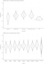
      Bookmark
          There is no author summary for this article yet. Authors can add summaries to their articles on ScienceOpen to make them more accessible to a non-specialist audience.

          Abstract

          Although previous studies have highlighted associations of cannabis use with cognition and brain morphometry, critical questions remain with regard to the association between cannabis use and brain structural and functional connectivity. In a cross‐sectional community sample of 205 African Americans (age 18–70) we tested for associations of cannabis use disorder (CUD, n = 57) with multi‐domain cognitive measures and structural, diffusion, and resting state brain‐imaging phenotypes. Post hoc model evidence was computed with Bayes factors (BF) and posterior probabilities of association (PPA) to account for multiple testing. General cognitive functioning, verbal intelligence, verbal memory, working memory, and motor speed were lower in the CUD group compared with non‐users ( p < .011; 1.9 < BF < 3,217). CUD was associated with altered functional connectivity in a network comprising the motor‐hand region in the superior parietal gyri and the anterior insula ( p < .04). These differences were not explained by alcohol, other drug use, or education. No associations with CUD were observed in cortical thickness, cortical surface area, subcortical or cerebellar volumes (0.12 < BF < 1.5), or graph‐theoretical metrics of resting state connectivity (PPA < 0.01). In a large sample collected irrespective of cannabis used to minimize recruitment bias, we confirm the literature on poorer cognitive functioning in CUD, and an absence of volumetric brain differences between CUD and non‐CUD. We did not find evidence for or against a disruption of structural connectivity, whereas we did find localized resting state functional dysconnectivity in CUD. There was sufficient proof, however, that organization of functional connectivity as determined via graph metrics does not differ between CUD and non‐user group.

          Abstract

          In a large sample collected irrespective of cannabis used to minimize recruitment bias, we confirm the literature on poorer cognitive functioning in cannabis use disorder (CUD), and an absence of volumetric brain differences between CUD and non‐CUD. A disruption of structural connectivity remains equivocal. We find localized resting state functional dysconnectivity in CUD, and sufficient proof that organization of functional connectivity as determined via graph metrics does not differ between CUD and non‐users.

          Related collections

          Most cited references92

          • Record: found
          • Abstract: found
          • Article: not found

          An automated labeling system for subdividing the human cerebral cortex on MRI scans into gyral based regions of interest.

          In this study, we have assessed the validity and reliability of an automated labeling system that we have developed for subdividing the human cerebral cortex on magnetic resonance images into gyral based regions of interest (ROIs). Using a dataset of 40 MRI scans we manually identified 34 cortical ROIs in each of the individual hemispheres. This information was then encoded in the form of an atlas that was utilized to automatically label ROIs. To examine the validity, as well as the intra- and inter-rater reliability of the automated system, we used both intraclass correlation coefficients (ICC), and a new method known as mean distance maps, to assess the degree of mismatch between the manual and the automated sets of ROIs. When compared with the manual ROIs, the automated ROIs were highly accurate, with an average ICC of 0.835 across all of the ROIs, and a mean distance error of less than 1 mm. Intra- and inter-rater comparisons yielded little to no difference between the sets of ROIs. These findings suggest that the automated method we have developed for subdividing the human cerebral cortex into standard gyral-based neuroanatomical regions is both anatomically valid and reliable. This method may be useful for both morphometric and functional studies of the cerebral cortex as well as for clinical investigations aimed at tracking the evolution of disease-induced changes over time, including clinical trials in which MRI-based measures are used to examine response to treatment.
            Bookmark
            • Record: found
            • Abstract: found
            • Article: not found

            FreeSurfer.

            FreeSurfer is a suite of tools for the analysis of neuroimaging data that provides an array of algorithms to quantify the functional, connectional and structural properties of the human brain. It has evolved from a package primarily aimed at generating surface representations of the cerebral cortex into one that automatically creates models of most macroscopically visible structures in the human brain given any reasonable T1-weighted input image. It is freely available, runs on a wide variety of hardware and software platforms, and is open source. Copyright © 2012 Elsevier Inc. All rights reserved.
              Bookmark
              • Record: found
              • Abstract: found
              • Article: not found

              Advances in functional and structural MR image analysis and implementation as FSL.

              The techniques available for the interrogation and analysis of neuroimaging data have a large influence in determining the flexibility, sensitivity, and scope of neuroimaging experiments. The development of such methodologies has allowed investigators to address scientific questions that could not previously be answered and, as such, has become an important research area in its own right. In this paper, we present a review of the research carried out by the Analysis Group at the Oxford Centre for Functional MRI of the Brain (FMRIB). This research has focussed on the development of new methodologies for the analysis of both structural and functional magnetic resonance imaging data. The majority of the research laid out in this paper has been implemented as freely available software tools within FMRIB's Software Library (FSL).
                Bookmark

                Author and article information

                Contributors
                maria.koenis@yale.edu
                Journal
                Hum Brain Mapp
                Hum Brain Mapp
                10.1002/(ISSN)1097-0193
                HBM
                Human Brain Mapping
                John Wiley & Sons, Inc. (Hoboken, USA )
                1065-9471
                1097-0193
                19 December 2020
                15 April 2021
                : 42
                : 6 ( doiID: 10.1002/hbm.v42.6 )
                : 1727-1741
                Affiliations
                [ 1 ] Department of Psychiatry School of Medicine, Yale University New Haven Connecticut USA
                [ 2 ] Olin Neuropsychiatry Research Center Institute of Living Hartford Connecticut USA
                [ 3 ] Department of Psychology Stanford University Stanford California USA
                [ 4 ] Department of Psychiatry Boston Children's Hospital & Harvard Medical School Boston Massachusetts USA
                [ 5 ] Department of Psychiatry Icahn School of Medicine at Mount Sinai New York New York USA
                [ 6 ] Department of Human Genetics, and South Texas Diabetes and Obesity Institute School of Medicine, University of Texas Rio Grande Valley Brownsville Texas USA
                [ 7 ] Department of Neuroscience Yale University New Haven Connecticut USA
                Author notes
                [*] [* ] Correspondence

                Marinka M. G. Koenis, Olin Neuropsychiatry Research Center, Institute of Living, 200 Retreat Ave, Hartford, CT 06106.

                Email: maria.koenis@ 123456yale.edu

                Author information
                https://orcid.org/0000-0002-3859-3847
                https://orcid.org/0000-0002-3403-6349
                https://orcid.org/0000-0003-4120-0474
                https://orcid.org/0000-0002-3210-6470
                https://orcid.org/0000-0001-9622-5420
                https://orcid.org/0000-0002-4749-6977
                Article
                HBM25324
                10.1002/hbm.25324
                7978126
                33340172
                97d32129-422c-4334-a0d0-0ef79d348845
                © 2020 The Authors. Human Brain Mapping published by Wiley Periodicals LLC.

                This is an open access article under the terms of the http://creativecommons.org/licenses/by/4.0/ License, which permits use, distribution and reproduction in any medium, provided the original work is properly cited.

                History
                : 31 August 2020
                : 11 February 2020
                : 10 December 2020
                Page count
                Figures: 3, Tables: 4, Pages: 15, Words: 12742
                Funding
                Funded by: National Institute on Drug Abuse , open-funder-registry 10.13039/100000026;
                Award ID: 1R01DA038807
                Funded by: National Institute of Mental Health , open-funder-registry 10.13039/100000025;
                Award ID: R01 MH106324
                Categories
                Research Article
                Research Articles
                Custom metadata
                2.0
                15 April 2021
                Converter:WILEY_ML3GV2_TO_JATSPMC version:6.0.0 mode:remove_FC converted:19.03.2021

                Neurology
                cognition,dti,marijuana,morphometry,resting state,white matter
                Neurology
                cognition, dti, marijuana, morphometry, resting state, white matter

                Comments

                Comment on this article