48
views
0
recommends
+1 Recommend
0 collections
    0
    shares
      • Record: found
      • Abstract: found
      • Article: found
      Is Open Access

      Early Activation of STAT3 Regulates Reactive Astrogliosis Induced by Diverse Forms of Neurotoxicity

      research-article

      Read this article at

      Bookmark
          There is no author summary for this article yet. Authors can add summaries to their articles on ScienceOpen to make them more accessible to a non-specialist audience.

          Abstract

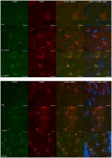
          Astrogliosis, a cellular response characterized by astrocytic hypertrophy and accumulation of GFAP, is a hallmark of all types of central nervous system (CNS) injuries. Potential signaling mechanisms driving the conversion of astrocytes into “reactive” phenotypes differ with respect to the injury models employed and can be complicated by factors such as disruption of the blood-brain barrier (BBB). As denervation tools, neurotoxicants have the advantage of selective targeting of brain regions and cell types, often with sparing of the BBB. Previously, we found that neuroinflammation and activation of the JAK2-STAT3 pathway in astrocytes precedes up regulation of GFAP in the MPTP mouse model of dopaminergic neurotoxicity. Here we show that multiple mechanistically distinct mouse models of neurotoxicity (MPTP, AMP, METH, MDA, MDMA, KA, TMT) engender the same neuroinflammatory and STAT3 activation responses in specific regions of the brain targeted by each neurotoxicant. The STAT3 effects seen for TMT in the mouse could be generalized to the rat, demonstrating cross-species validity for STAT3 activation. Pharmacological antagonists of the neurotoxic effects blocked neuroinflammatory responses, pSTAT3 tyr705 and GFAP induction, indicating that damage to neuronal targets instigated astrogliosis. Selective deletion of STAT3 from astrocytes in STAT3 conditional knockout mice markedly attenuated MPTP-induced astrogliosis. Monitoring STAT3 translocation in GFAP-positive cells indicated that effects of MPTP, METH and KA on pSTAT3 tyr705 were localized to astrocytes. These findings strongly implicate the STAT3 pathway in astrocytes as a broadly triggered signaling pathway for astrogliosis. We also observed, however, that the acute neuroinflammatory response to the known inflammogen, LPS, can activate STAT3 in CNS tissue without inducing classical signs of astrogliosis. Thus, acute phase neuroinflammatory responses and neurotoxicity-induced astrogliosis both signal through STAT3 but appear to do so through different modules, perhaps localized to different cell types.

          Related collections

          Most cited references52

          • Record: found
          • Abstract: found
          • Article: not found

          Systemic LPS causes chronic neuroinflammation and progressive neurodegeneration.

          Inflammation is implicated in the progressive nature of neurodegenerative diseases, such as Parkinson's disease, but the mechanisms are poorly understood. A single systemic lipopolysaccharide (LPS, 5 mg/kg, i.p.) or tumor necrosis factor alpha (TNFalpha, 0.25 mg/kg, i.p.) injection was administered in adult wild-type mice and in mice lacking TNFalpha receptors (TNF R1/R2(-/-)) to discern the mechanisms of inflammation transfer from the periphery to the brain and the neurodegenerative consequences. Systemic LPS administration resulted in rapid brain TNFalpha increase that remained elevated for 10 months, while peripheral TNFalpha (serum and liver) had subsided by 9 h (serum) and 1 week (liver). Systemic TNFalpha and LPS administration activated microglia and increased expression of brain pro-inflammatory factors (i.e., TNFalpha, MCP-1, IL-1beta, and NF-kappaB p65) in wild-type mice, but not in TNF R1/R2(-/-) mice. Further, LPS reduced the number of tyrosine hydroxylase-immunoreactive neurons in the substantia nigra (SN) by 23% at 7-months post-treatment, which progressed to 47% at 10 months. Together, these data demonstrate that through TNFalpha, peripheral inflammation in adult animals can: (1) activate brain microglia to produce chronically elevated pro-inflammatory factors; (2) induce delayed and progressive loss of DA neurons in the SN. These findings provide valuable insight into the potential pathogenesis and self-propelling nature of Parkinson's disease. (c) 2007 Wiley-Liss, Inc.
            Bookmark
            • Record: found
            • Abstract: found
            • Article: not found

            The JAK-STAT signaling pathway: input and output integration.

            Universal and essential to cytokine receptor signaling, the JAK-STAT pathway is one of the best understood signal transduction cascades. Almost 40 cytokine receptors signal through combinations of four JAK and seven STAT family members, suggesting commonality across the JAK-STAT signaling system. Despite intense study, there remain substantial gaps in understanding how the cascades are activated and regulated. Using the examples of the IL-6 and IL-10 receptors, I will discuss how diverse outcomes in gene expression result from regulatory events that effect the JAK1-STAT3 pathway, common to both receptors. I also consider receptor preferences by different STATs and interpretive problems in the use of STAT-deficient cells and mice. Finally, I consider how the suppressor of cytokine signaling (SOCS) proteins regulate the quality and quantity of STAT signals from cytokine receptors. New data suggests that SOCS proteins introduce additional diversity into the JAK-STAT pathway by adjusting the output of activated STATs that alters downstream gene activation.
              Bookmark
              • Record: found
              • Abstract: found
              • Article: not found

              Glial scar borders are formed by newly proliferated, elongated astrocytes that interact to corral inflammatory and fibrotic cells via STAT3-dependent mechanisms after spinal cord injury.

              Astroglial scars surround damaged tissue after trauma, stroke, infection, or autoimmune inflammation in the CNS. They are essential for wound repair, but also interfere with axonal regrowth. A better understanding of the cellular mechanisms, regulation, and functions of astroglial scar formation is fundamental to developing safe interventions for many CNS disorders. We used wild-type and transgenic mice to quantify and dissect these parameters. Adjacent to crush spinal cord injury (SCI), reactive astrocytes exhibited heterogeneous phenotypes as regards proliferation, morphology, and chemistry, which all varied with distance from lesions. Mature scar borders at 14 d after SCI consisted primarily of newly proliferated astroglia with elongated cell processes that surrounded large and small clusters of inflammatory, fibrotic, and other cells. During scar formation from 5 to 14 d after SCI, cell processes deriving from different astroglia associated into overlapping bundles that quantifiably reoriented and organized into dense mesh-like arrangements. Selective deletion of STAT3 from astroglia quantifiably disrupted the organization of elongated astroglia into scar borders, and caused a failure of astroglia to surround inflammatory cells, resulting in increased spread of these cells and neuronal loss. In cocultures, wild-type astroglia spontaneously corralled inflammatory or fibromeningeal cells into segregated clusters, whereas STAT3-deficient astroglia failed to do so. These findings demonstrate heterogeneity of reactive astroglia and show that scar borders are formed by newly proliferated, elongated astroglia, which organize via STAT3-dependent mechanisms to corral inflammatory and fibrotic cells into discrete areas separated from adjacent tissue that contains viable neurons.
                Bookmark

                Author and article information

                Contributors
                Role: Editor
                Journal
                PLoS One
                PLoS ONE
                plos
                plosone
                PLoS ONE
                Public Library of Science (San Francisco, USA )
                1932-6203
                2014
                15 July 2014
                : 9
                : 7
                : e102003
                Affiliations
                [1 ]Centers for Disease Control and Prevention, National Institute for Occupational Safety and Health, Morgantown, West Virginia, United States of America
                [2 ]School of Pharmacy, West Virginia University, Morgantown, West Virginia, United States of America
                [3 ]David Geffen School of Medicine, University of California Los Angeles, Los Angeles, California, United States of America
                University of Nebraska Medical Center, United States of America
                Author notes

                Competing Interests: The authors have declared that no competing interests exist.

                Conceived and designed the experiments: JPO DBM MVS KAK. Performed the experiments: KAK RLV JPO. Analyzed the data: KAK JPO DBM. Contributed reagents/materials/analysis tools: KAK RLV MVS JPO. Contributed to the writing of the manuscript: KAK JPO DBM MVS.

                Article
                PONE-D-14-12852
                10.1371/journal.pone.0102003
                4098997
                25025494
                97db7299-8c3f-4134-9122-5b056022d116
                Copyright @ 2014

                This is an open-access article, free of all copyright, and may be freely reproduced, distributed, transmitted, modified, built upon, or otherwise used by anyone for any lawful purpose. The work is made available under the Creative Commons CC0 public domain dedication.

                History
                : 21 March 2014
                : 13 June 2014
                Page count
                Pages: 17
                Funding
                This study was funded by intramural funds from the Centers for Disease Control and Prevention, National Institute for Occupational Safety and Health. The funders had no role in study design, data collection and analysis, decision to publish, or preparation of the manuscript.
                Categories
                Research Article
                Biology and Life Sciences
                Cell Biology
                Cellular Types
                Animal Cells
                Glial Cells
                Macroglial Cells
                Astrocytes
                Neuroscience
                Molecular Neuroscience
                Toxicology
                Toxicity
                Custom metadata
                The authors confirm that all data underlying the findings are fully available without restriction. All relevant data are within the paper. All data are available upon request.

                Uncategorized
                Uncategorized

                Comments

                Comment on this article