3
views
0
recommends
+1 Recommend
0 collections
    0
    shares
      • Record: found
      • Abstract: found
      • Article: found
      Is Open Access

      Anatomic Posterolateral Corner Reconstruction With Autografts

      brief-report

      Read this article at

      Bookmark
          There is no author summary for this article yet. Authors can add summaries to their articles on ScienceOpen to make them more accessible to a non-specialist audience.

          Abstract

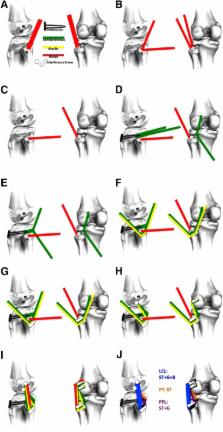
          Anatomic posterolateral corner reconstruction reproduces 3 main structures: the lateral collateral ligament, the popliteofibular ligament, and the popliteus tendon. The LaPrade technique reproduces all 3 main stabilizers. However, it requires a long graft, limiting its indication to clinical settings in which allograft tissue is available. We propose a surgical procedure that is a modification of the LaPrade technique using the same tunnel placement, hamstring autografts, and biceps augmentation when necessary. It relies on artificial graft lengthening provided by the loop of the suspensory fixation device fixed at the anterior tibial cortex. The final reconstruction reproduces the popliteus tendon with the bulkiest end of the semitendinosus; the popliteofibular ligament with a strand of the semitendinosus and a strand of the gracilis; and the lateral collateral ligament with a strand of the semitendinosus and a strand of the gracilis, which can also be augmented with a biceps strip.

          Related collections

          Most cited references12

          • Record: found
          • Abstract: found
          • Article: not found

          An analysis of an anatomical posterolateral knee reconstruction: an in vitro biomechanical study and development of a surgical technique.

          To date, no surgical technique to treat posterolateral knee instability anatomically reconstructs the 3 major static stabilizing structures of the posterolateral knee: the fibular collateral ligament, the popliteus tendon, and the popliteofibular ligament. Static varus and external rotatory stability would be restored to the reconstructed knee with a posterolateral knee injury. The anatomical locations of the original fibular collateral ligament, popliteus tendon, and popliteofibular ligament were reconstructed using a 2-graft technique. Ten cadaveric specimens were tested in 3 states: intact knee, knee with the 3 structures cut to simulate a grade III injury, and the reconstructed knee. For the varus loading tests, joint stability was significantly improved by the posterolateral reconstruction compared to the cut state at 0 degrees, 30 degrees, 60 degrees, and 90 degrees of flexion. There were no significant differences between the intact and reconstructed knees at 0 degrees, 60 degrees, and 90 degrees for varus translation. For the external rotation torque tests, external rotation was significantly higher for the cut state than for the intact or reconstructed posterolateral knee. There was no significant difference in external rotation between the intact and reconstructed posterolateral knees at any flexion angle. This 2-graft technique to reconstruct the primary static stabilizers of the posterolateral knee restored static stability, as measured by joint translation in response to varus loading and external rotation torque, to knees with grade III posterolateral injuries.
            Bookmark
            • Record: found
            • Abstract: found
            • Article: not found

            Posterolateral Corner of the Knee: Current Concepts.

            Injuries to the posterolateral corner (PLC) comprise a significant portion of knee ligament injuries. A high index of suspicion is necessary when evaluating the injured knee to detect these sometimes occult injuries. Moreover, a thorough physical examination and a comprehensive review of radiographic studies are necessary to identify these injuries. In this sense, stress radiographs can help to objectively determine the extent of these lesions. Non-operative and operative treatment options have been reported depending on the extent of the injury. Complete PLC lesions rarely heal with non-operative treatment, and are therefore most often treated surgically. The purpose of this article was to review the anatomy and clinically relevant biomechanics, diagnosis algorithms, treatment and rehabilitation protocols for PLC injuries.
              Bookmark
              • Record: found
              • Abstract: found
              • Article: not found
              Is Open Access

              Anatomic Posterolateral Corner Reconstruction

              Posterolateral corner injuries represent a complex injury pattern, with damage to important coronal and rotatory stabilizers of the knee. These lesions commonly occur in association with other ligament injuries, making decisions regarding treatment challenging. Grade III posterolateral corner injuries result in significant instability and have poor outcomes when treated nonoperatively. As a result, reconstruction is advocated. A thorough knowledge of the anatomy is essential for surgical treatment of this pathology. The following technical note provides a diagnostic approach, postoperative management, and details of a technique for anatomic reconstruction of the 3 main static stabilizers of the posterolateral corner of the knee.
                Bookmark

                Author and article information

                Contributors
                Journal
                Arthrosc Tech
                Arthrosc Tech
                Arthroscopy Techniques
                Elsevier
                2212-6287
                08 January 2018
                February 2018
                08 January 2018
                : 7
                : 2
                : e89-e95
                Affiliations
                [a ]Department of Orthopaedics and Traumatology, Escola Paulista de Medicina, Federal University of São Paulo, São Paulo, Brazil
                [b ]Hospital Israelita Albert Einstein, São Paulo, Brazil
                [c ]Knee Institute, Hospital do Coração (HCor), São Paulo, Brazil
                [d ]Associação de Assistência à Criança Deficiente, São Paulo, Brazil
                Author notes
                []Address correspondence to Carlos Eduardo Franciozi, M.D., Ph.D., Department of Orthopaedics and Traumatology, Escola Paulista de Medicina, Federal University of São Paulo, Rua Borges Lagoa, 783, Fifth Floor, Vila Clementino, São Paulo, Brazil 04038-032.Department of Orthopaedics and TraumatologyEscola Paulista de MedicinaFederal University of São PauloRua Borges Lagoa783, Fifth FloorVila ClementinoSão Paulo04038-032Brazil cacarlos66@ 123456hotmail.com
                Article
                S2212-6287(17)30321-3
                10.1016/j.eats.2017.08.053
                5869793
                29593980
                996799f0-bce4-4bf0-9384-a05209098649
                © 2017 by the Arthroscopy Association of North America. Published by Elsevier.

                This is an open access article under the CC BY-NC-ND license (http://creativecommons.org/licenses/by-nc-nd/4.0/).

                History
                : 11 April 2017
                : 10 August 2017
                Categories
                Technical Note

                Comments

                Comment on this article