3
views
0
recommends
+1 Recommend
0 collections
    0
    shares
      • Record: found
      • Abstract: found
      • Article: found
      Is Open Access

      M2 macrophage-induced lncRNA PCAT6 facilitates tumorigenesis and angiogenesis of triple-negative breast cancer through modulation of VEGFR2

      research-article

      Read this article at

      Bookmark
          There is no author summary for this article yet. Authors can add summaries to their articles on ScienceOpen to make them more accessible to a non-specialist audience.

          Abstract

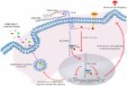
          As a common female malignancy, triple-negative breast cancer (TNBC) is the most malignant subtype of breast cancers (BC). This study further studied the role of long noncoding RNA (lncRNA) prostate cancer-associated transcript 6 (PCAT6) in TNBC. Functional assays, including EdU, wound healing, transwell, and immunofluorescence staining, revealed the effect of PCAT6 on cell proliferation, migration, and EMT process. The tube-formation assay disclosed the function of PCAT6 on angiogenesis. In vivo assays were also established to explore the impact of PCAT6 on tumor growth and microangiogenesis. The results revealed that PCAT6 boosted TNBC cell proliferation, migration, and angiogenesis both in vitro and in vivo. Then, this study unveiled that M2 macrophage secreted VEGF to stimulate the upregulation of PCAT6, thus promoting angiogenesis in TNBC. Next, through bioinformatics analysis and mechanism assays, we identified that PCAT6 positively regulated VEGFR2 expression via ceRNA pattern and then participated in VEGFR/AKT/mTOR signaling pathway to accelerate angiogenesis. Moreover, PCAT6 bound USP14, a deubiquitinase, to induce the deubiquitination of VEGFR2. On the whole, M2 macrophage-induced upregulation of PCAT6 facilitates TNBC tumorigenesis through modulation of VEGFR2 expression via ceRNA and deubiquitination patterns.

          Related collections

          Most cited references25

          • Record: found
          • Abstract: found
          • Article: not found

          High numbers of macrophages, especially M2-like (CD163-positive), correlate with hyaluronan accumulation and poor outcome in breast cancer.

          High amounts of tumour-associated macrophages (TAMs) and hyaluronan (HA) correlate with tumour aggressiveness in breast cancer, but the relationship between these parameters is unclear. The aim of this study was to assay the numbers of TAMs in 278 human breast cancer cases, and their correlations with HA-related factors, clinical variables, and outcome.
            Bookmark
            • Record: found
            • Abstract: found
            • Article: not found

            Macrophage Polarization: Anti-Cancer Strategies to Target Tumor-Associated Macrophage in Breast Cancer.

            Tumor-associated macrophages (TAMs) are the most abundant inflammatory cells and orchestrate different stages of breast cancer development. TAMs participate in the tumor angiogenesis, matrix remodeling, invasion, immunosuppression, metastasis, and chemoresistance in breast cancer. Several clinical studies indicate the association between the high influx of TAMs in tumor with poor prognosis in hepatocellular, ovarian, cervical, and breast cancer. Previously developed hypotheses have proposed that TAMs participate in antitumor responses of the body, while recently many clinical and experimental studies have revealed that TAMs in tumor microenvironment predominantly resemble with M2-like polarized macrophages and produce a high amount of anti-inflammatory factors which are directly responsible for the development of tumor. Various studies have shown that TAMs in tumor either enhance or antagonize the anti-tumor efficacy of cytotoxic agents, antibodies-targeting cancer cells, and therapeutic agents depending on the nature of treatment. Thereby, multiple roles of TAMs suggests that it is very important to develop novel therapeutic strategies to target TAMs in breast tumor. In this review, we have discussed the functional role of TAMs in breast cancer and summarized available recent advances potential therapeutic strategies that effectively target to TAMs cells. J. Cell. Biochem. 118: 2484-2501, 2017. © 2017 Wiley Periodicals, Inc.
              Bookmark
              • Record: found
              • Abstract: found
              • Article: found
              Is Open Access

              Hyaluronan, Inflammation, and Breast Cancer Progression

              Breast cancer-induced inflammation in the tumor reactive stroma supports invasion and malignant progression and is contributed to by a variety of host cells including macrophages and fibroblasts. Inflammation appears to be initiated by tumor cells and surrounding host fibroblasts that secrete pro-inflammatory cytokines and chemokines and remodel the extracellular matrix (ECM) to create a pro-inflammatory “cancerized” or tumor reactive microenvironment that supports tumor expansion and invasion. The tissue polysaccharide hyaluronan (HA) is an example of an ECM component within the cancerized microenvironment that promotes breast cancer progression. Like many ECM molecules, the function of native high-molecular weight HA is altered by fragmentation, which is promoted by oxygen/nitrogen free radicals and release of hyaluronidases within the tumor microenvironment. HA fragments are pro-inflammatory and activate signaling pathways that promote survival, migration, and invasion within both tumor and host cells through binding to HA receptors such as CD44 and RHAMM/HMMR. In breast cancer, elevated HA in the peri-tumor stroma and increased HA receptor expression are prognostic for poor outcome and are associated with disease recurrence. This review addresses the critical issues regarding tumor-induced inflammation and its role in breast cancer progression focusing specifically on the changes in HA metabolism within tumor reactive stroma as a key factor in malignant progression.
                Bookmark

                Author and article information

                Contributors
                huting0709@hust.edu.cn
                wdyxywq@hust.edu.cn
                Journal
                Cell Death Dis
                Cell Death Dis
                Cell Death & Disease
                Nature Publishing Group UK (London )
                2041-4889
                9 September 2020
                9 September 2020
                September 2020
                : 11
                : 9
                : 728
                Affiliations
                [1 ]GRID grid.33199.31, ISNI 0000 0004 0368 7223, Department of Breast and Thyroid Surgery, Union Hospital, Tongji Medical College, , Huazhong University of Science and Technology, ; 430022 Wuhan, Hubei China
                [2 ]GRID grid.33199.31, ISNI 0000 0004 0368 7223, Department of Orthopaedic, Union Hospital, Tongji Medical College, , Huazhong University of Science and Technology, ; 430022 Wuhan, Hubei China
                [3 ]GRID grid.33199.31, ISNI 0000 0004 0368 7223, Cancer Center, Union Hospital, Tongji Medical College, , Huazhong University of Science and Technology, ; 430022 Wuhan, Hubei China
                Article
                2926
                10.1038/s41419-020-02926-8
                7481779
                31911576
                99f18703-138e-405f-9c8a-3013e35e1100
                © The Author(s) 2020

                Open Access This article is licensed under a Creative Commons Attribution 4.0 International License, which permits use, sharing, adaptation, distribution and reproduction in any medium or format, as long as you give appropriate credit to the original author(s) and the source, provide a link to the Creative Commons license, and indicate if changes were made. The images or other third party material in this article are included in the article’s Creative Commons license, unless indicated otherwise in a credit line to the material. If material is not included in the article’s Creative Commons license and your intended use is not permitted by statutory regulation or exceeds the permitted use, you will need to obtain permission directly from the copyright holder. To view a copy of this license, visit http://creativecommons.org/licenses/by/4.0/.

                History
                : 12 February 2020
                : 16 July 2020
                : 16 July 2020
                Funding
                Funded by: This project was supported by grants from the National Natural Science Foundation of China (No. 81703032).
                Categories
                Article
                Custom metadata
                © The Author(s) 2020

                Cell biology
                breast cancer,cell biology
                Cell biology
                breast cancer, cell biology

                Comments

                Comment on this article