20
views
0
recommends
+1 Recommend
0 collections
    0
    shares
      • Record: found
      • Abstract: found
      • Article: found
      Is Open Access

      Association of Abdominal Aortic Calcification with Lifestyle and Risk Factors of Cardiovascular Disease

      research-article

      Read this article at

      Bookmark
          There is no author summary for this article yet. Authors can add summaries to their articles on ScienceOpen to make them more accessible to a non-specialist audience.

          Abstract

          Background

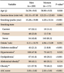
          Abdominal aortic calcification (AAC) is a marker of subclinical atherosclerotic disease and an independent predictor of subsequent vascular morbidity and mortality. This study was conducted to investigate the association of AAC with lifestyle and risk factors of cardiovascular disease.

          Methods

          The results of the abdominal computed tomography of 380 patients who visited Chungnam National University Hospital for a health checkup from January 1, 2008 to December 31, 2009 were reviewed. A six-point scale was used in grading the overall severity of the calcification in three areas of the abdominal aorta, including the area superior to the renal artery, the upper-half area inferior to the renal artery, and the lower-half area inferior to the renal artery, in addition to the common iliac artery. The association of the AAC severity with the age, lifestyle factors, and risk factors of cardiovascular disease was analyzed via multiple linear regression analysis.

          Results

          In the male subjects, the age, presence of dyslipidemia and smoking were positively related to AAC, but exercising was negatively related to AAC (total R 2 = 0.563). In the female subjects, the age and presence of diabetes mellitus, hypertension, and dyslipidemia were positively related to AAC, but exercising was negatively related to AAC (total R 2 = 0.547).

          Conclusion

          AAC was related to both the male and female subjects' age, presence of dyslipidemia, and exercising, to smoking in the male subjects and to the presence of diabetes mellitus and hypertension in the female subjects.

          Related collections

          Most cited references25

          • Record: found
          • Abstract: not found
          • Article: not found

          Report of the Expert Committee on the Diagnosis and Classification of Diabetes Mellitus

          (2002)
            Bookmark
            • Record: found
            • Abstract: found
            • Article: not found

            New indices to classify location, severity and progression of calcific lesions in the abdominal aorta: a 25-year follow-up study.

            L Kauppila (1997)
            The purpose of the present study was to assess the location, severity and progression of radiopaque lumbar aortic calcifications and to evaluate the utility of summary scores of lumbar calcification in a population-based cohort. Lateral lumbar films, obtained in 617 Framingham heart study participants, were analysed for the presence of abdominal aortic wall calcification in the region corresponding to the first through fourth lumbar vertebrae. The severity of the anterior and posterior aortic calcification were graded individually on a 0-3 scale for each lumbar segment and the results were summarized to develop four different composite scores: (1) affected segments score (range 0-4); (2) anterior and posterior affected score (range 0-8); and (3) antero-posterior severity score (range 0-24). The prevalence of aortic calcification was 37% in men and 27% in women at baseline and 86% in both genders at the follow-up exam 25 years later. During the follow-up interval, the mean of the affected segments score increased from 0.7 in men (0.5 in women) to 2.7 (2.8 in women), the mean of the anterior and posterior affected score from 1.2 (0.8 in women) (P = 0.012 for difference between genders) and the mean of the antero-posterior severity score increased from 1.5 (1.3 in women) to 9.3 (10.3 in women). The antero-posterior severity score offered a slight advantage over other composite scores and had the highest inter-rater intra-class correlations. In summary, lumbar aortic calcification can be graded and composite summary scores are reproducible. This technique appears to provide a simple, low cost assessment of subclinical vascular disease.
              Bookmark
              • Record: found
              • Abstract: found
              • Article: not found

              Coronary artery calcium area by electron-beam computed tomography and coronary atherosclerotic plaque area. A histopathologic correlative study.

              Coronary calcium identified by electron-beam computed tomography (EBCT) correlates poorly with luminal atherosclerotic narrowing, but calcium, an intimate part of coronary plaque, may be more directly related to atheromatous plaque area. Thirty-eight coronary arteries from 13 autopsy hearts were dissected, straightened, and scanned with EBCT in 3-mm contiguous increments. Coronary calcium area was defined as one or more pixels with a density > 130 Hounsfield units (0.18 mm2/pixel). Each artery was divided into corresponding 3-mm segments, representative histological sections were stained, and atherosclerotic plaque area per segment (mm2) was quantified. Coronary artery calcium and coronary artery plaque areas were correlated for the hearts as a whole, for individual coronary arteries, and for individual coronary artery segments. The sums of histological plaque areas versus the sums of calcium areas were highly correlated for each heart and for each coronary artery. However, coronary plaque area was on the order of five times greater than calcium area. Furthermore, minimal diffuse segmental coronary plaque could be present despite the absence of coronary calcium detectable by EBCT. This histopathologic study confirms an intimate relation between whole heart, coronary artery, and segmental coronary atherosclerotic plaque area and EBCT coronary calcium area but suggests that there is a threshold value for plaque area below which coronary calcium is either absent or not detectable by this methodology.
                Bookmark

                Author and article information

                Journal
                Korean J Fam Med
                Korean J Fam Med
                KJFM
                Korean Journal of Family Medicine
                The Korean Academy of Family Medicine
                2005-6443
                2092-6715
                May 2013
                24 May 2013
                : 34
                : 3
                : 213-220
                Affiliations
                Department of Family Medicine, Research Institute for Medical Sciences, Chungnam National University School of Medicine, Daejeon, Korea.
                Author notes
                Corresponding Author: Jong Sung Kim. Tel: +82-42-280-8172, Fax: +82-42-280-7879, jskim@ 123456cnuh.co.kr
                Article
                10.4082/kjfm.2013.34.3.213
                3667229
                23730489
                9bac4744-31b1-4606-89b2-99d8d2112b68
                Copyright © 2013 The Korean Academy of Family Medicine

                This is an open-access article distributed under the terms of the Creative Commons Attribution Non-Commercial License ( http://creativecommons.org/licenses/by-nc/3.0/) which permits unrestricted noncommercial use, distribution, and reproduction in any medium, provided the original work is properly cited.

                History
                : 31 August 2011
                : 28 February 2013
                Categories
                Original Article

                Medicine
                aorta,calcification,atherosclerosis,life style
                Medicine
                aorta, calcification, atherosclerosis, life style

                Comments

                Comment on this article