28
views
0
recommends
+1 Recommend
0 collections
    0
    shares
      • Record: found
      • Abstract: found
      • Article: found
      Is Open Access

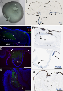
      The expression of Wnt2b in the optic cup lip requires a border between the pigmented and nonpigmented epithelium

      research-article
      ,
      Molecular Vision
      Molecular Vision

      Read this article at

      Bookmark
          There is no author summary for this article yet. Authors can add summaries to their articles on ScienceOpen to make them more accessible to a non-specialist audience.

          Abstract

          Purpose

          Wnt2b is normally expressed at the optic cup lip and is implicated in ciliary body induction. The lens has often been considered an organizer for the anterior eye, but recent studies demonstrate that the anterior cell fates are correctly specified in the absence of the lens. This study uses Wnt2b as a marker to reveal the mechanism behind the specification of the anterior domain of the optic cup.

          Methods

          Developing chick embryos were used as a model system. Eyes were microsurgically manipulated to assess the role of the lens in the development of the anterior optic cup. Eyes were molecularly manipulated, using fibroblast growth factor expressing replication-incompetent retrovirus, introduced into the retinal pigmented epithelium (RPE) domain. Ectopic fibroblast growth factor transformed the RPE into nonpigmented epithelium (NPE; ciliary body). As the virus does not spread, discrete borders between RPE and NPE were experimentally created. Wnt2b expression was assessed after surgical and molecular manipulation.

          Results

          Contrary to expectations, we found that the lens is not able to induce Wnt2b expression in optic cup tissue: When the optic cup lip is experimentally misspecified such that it no longer contains the juxtaposition of pigmented and nonpigmented tissue, Wnt2b is not expressed. In addition, if the prelens ectoderm is removed from the optic vesicle before morphogenesis, the resulting lensless optic cup expresses Wnt2b even though it was not in contact with lens tissue. We also show that ectopic lenses do not induce Wnt2b in optic cup tissue. The ciliary body/anterior eye domain is specified at the border of RPE and the NPE of the ciliary body. During development, this border is normally found at the optic cup lip. We can manipulate tissue specification using retroviral-mediated gene transfer, and create ectopic borders between nonpigmented and pigmented tissue. At such borders, Wnt2b is ectopically expressed in the absence of lens contact. Finally, we describe a role for the lens in maintenance of Wnt2b expression and demonstrate support for this in two ways: First, we show that if the lens is removed from the formed optic cup, endogenous Wnt2b expression is specifically lost from the optic cup lip; and second, we show that while ectopic Wnt2b expression is initially found in the majority of ectopic borders, as eye development proceeds ectopic expression is maintained only in those borders that are close to the lens.

          Conclusions

          Taken together, the results provide support for a model in which the anterior optic cup domain, as described in part by Wnt2b expression, is specified through the elaboration of a border within the optic neuroepithelium rather than through interactions with the surrounding environment.

          Related collections

          Most cited references54

          • Record: found
          • Abstract: not found
          • Article: not found

          A series of normal stages in the development of the chick embryo. 1951.

            Bookmark
            • Record: found
            • Abstract: found
            • Article: not found

            Beta-catenin controls differentiation of the retinal pigment epithelium in the mouse optic cup by regulating Mitf and Otx2 expression.

            The retinal pigment epithelium (RPE) consists of a monolayer of cuboidal, pigmented cells that is located between the retina and the choroid. The RPE is vital for growth and function of the vertebrate eye and improper development results in congenital defects, such as microphthalmia or anophthalmia, or a change of cell fate into neural retina called transdifferentiation. The transcription factors microphthalmia-associated transcription factor (Mitf) and orthodenticle homolog 2 (Otx2) are crucial for RPE development and function; however, very little is known about their regulation. Here, by using a Wnt-responsive reporter, we show that the Wnt/beta-catenin pathway is activated in the differentiating mouse RPE. Cre-mediated, RPE-specific disruption of beta-catenin after the onset of RPE specification causes severe defects, resulting in microphthalmia with coloboma, disturbed lamination, and mislocalization of adherens junction proteins. Upon beta-catenin deletion, the RPE transforms into a multilayered tissue in which the expression of Mitf and Otx2 is downregulated, while retina-specific gene expression is induced, which results in the transdifferentiation of RPE into retina. Chromatin immunoprecipitation (ChIP) and luciferase assays indicate that beta-catenin binds near to and activates potential TCF/LEF sites in the Mitf and Otx2 enhancers. We conclude that Wnt/beta-catenin signaling is required for differentiation of the RPE by directly regulating the expression of Mitf and Otx2. Our study is the first to show that an extracellular signaling pathway directly regulates the expression of RPE-specific genes such as Mitf and Otx2, and elucidates a new role for the Wnt/beta-catenin pathway in organ formation and development.
              Bookmark
              • Record: found
              • Abstract: found
              • Article: not found

              Central role for the lens in cave fish eye degeneration.

              Astyanax mexicanus is a teleost with eyed surface-dwelling and eyeless cave-dwelling forms. Eye formation is initiated in cave fish embryos, but the eye subsequently arrests and degenerates. The surface fish lens stimulates growth and development after transplantation into the cave fish optic cup, restoring optic tissues lost during cave fish evolution. Conversely, eye growth and development are retarded following transplantation of a surface fish lens into a cave fish optic cup or lens extirpation. These results show that evolutionary changes in an inductive signal from the lens are involved in cave fish eye degeneration.
                Bookmark

                Author and article information

                Journal
                Mol Vis
                MV
                Molecular Vision
                Molecular Vision
                1090-0535
                2010
                14 December 2010
                : 16
                : 2701-2717
                Affiliations
                [1]Department of Ophthalmology and Neurosurgery, University of California, San Francisco
                Author notes
                Correspondence to: Jeanette Hyer, Neurosurgery Department, University of California, San Francisco, Box 0112, 513 Parnassus Avenue, San Francisco, CA, 94143; Phone: (415) 515 4487; FAX: (415) 353 3907; email: jeanette.hyer@ 123456ucsf.edu
                Article
                289 2010MOLVIS0337
                3002962
                21179237
                9d7f6821-df9b-4222-90fd-08e86bfebd38
                Copyright © 2010 Molecular Vision.

                This is an open-access article distributed under the terms of the Creative Commons Attribution License, which permits unrestricted use, distribution, and reproduction in any medium, provided the original work is properly cited.

                History
                : 16 August 2010
                : 09 December 2010
                Categories
                Research Article
                Custom metadata
                Export to XML
                Hyer

                Vision sciences
                Vision sciences

                Comments

                Comment on this article