54
views
0
recommends
+1 Recommend
0 collections
    4
    shares
      • Record: found
      • Abstract: found
      • Article: found
      Is Open Access

      Respiratory Syncytial Virus Interferon Antagonist NS1 Protein Suppresses and Skews the Human T Lymphocyte Response

      research-article

      Read this article at

      Bookmark
          There is no author summary for this article yet. Authors can add summaries to their articles on ScienceOpen to make them more accessible to a non-specialist audience.

          Abstract

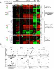
          We recently demonstrated that the respiratory syncytial virus (RSV) NS1 protein, an antagonist of host type I interferon (IFN-I) production and signaling, has a suppressive effect on the maturation of human dendritic cells (DC) that was only partly dependent on released IFN-I. Here we investigated whether NS1 affects the ability of DC to activate CD8+ and CD4+ T cells. Human DC were infected with RSV deletion mutants lacking the NS1 and/or NS2 genes and assayed for the ability to activate autologous T cells in vitro, which were analyzed by multi-color flow cytometry. Deletion of the NS1, but not NS2, protein resulted in three major effects: (i) an increased activation and proliferation of CD8+ T cells that express CD103, a tissue homing integrin that directs CD8+ T cells to mucosal epithelial cells of the respiratory tract and triggers cytolytic activity; (ii) an increased activation and proliferation of Th17 cells, which have recently been shown to have anti-viral effects and also indirectly attract neutrophils; and (iii) decreased activation of IL-4-producing CD4+ T cells - which are associated with enhanced RSV disease - and reduced proliferation of total CD4+ T cells. Except for total CD4+ T cell proliferation, none of the T cell effects appeared to be due to increased IFN-I signaling. In the infected DC, deletion of the NS1 and NS2 genes strongly up-regulated the expression of cytokines and other molecules involved in DC maturation. This was partly IFN-I-independent, and thus might account for the T cell effects. Taken together, these data demonstrate that the NS1 protein suppresses proliferation and activation of two of the protective cell populations (CD103+ CD8+ T cells and Th17 cells), and promotes proliferation and activation of Th2 cells that can enhance RSV disease.

          Author Summary

          Respiratory syncytial virus (RSV) is a leading cause of pediatric lower respiratory tract disease. RSV has two IFN-I antagonist proteins, NS1 and NS2. In this study, we infected primary human dendritic cells with recombinant RSV from which the NS1 and/or the NS2 genes were deleted, and evaluated effects on the proliferation of autologous T lymphocytes during co-culture in vitro. We found that NS1, but not NS2, has a suppressive effect on two cell populations, namely CD103+ CD8+ T cells and Th17 cells, which are known to protect against viral respiratory infections, and a stimulatory effect on Th2 cells, which are involved in enhanced disease caused by RSV. We also provide evidence that these effects are not due to suppressed IFN-I production or signaling in dendritic cells or T cells, and that they likely result from reduced maturation of dendritic cells caused by the NS1 protein.

          Related collections

          Most cited references75

          • Record: found
          • Abstract: found
          • Article: not found

          Type I interferons act directly on CD8 T cells to allow clonal expansion and memory formation in response to viral infection

          T cell expansion and memory formation are generally more effective when elicited by live organisms than by inactivated vaccines. Elucidation of the underlying mechanisms is important for vaccination and therapeutic strategies. We show that the massive expansion of antigen-specific CD8 T cells that occurs in response to viral infection is critically dependent on the direct action of type I interferons (IFN-Is) on CD8 T cells. By examining the response to infection with lymphocytic choriomeningitis virus using IFN-I receptor–deficient (IFN-IR0) and –sufficient CD8 T cells adoptively transferred into normal IFN-IR wild-type hosts, we show that the lack of direct CD8 T cell contact with IFN-I causes >99% reduction in their capacity to expand and generate memory cells. The diminished expansion of IFN-IR0 CD8 T cells was not caused by a defect in proliferation but by poor survival during the antigen-driven proliferation phase. Thus, IFN-IR signaling in CD8 T cells is critical for the generation of effector and memory cells in response to viral infection.
            Bookmark
            • Record: found
            • Abstract: found
            • Article: not found

            Induction and effector functions of T(H)17 cells.

            T helper (T(H)) cells constitute an important arm of the adaptive immune system because they coordinate defence against specific pathogens, and their unique cytokines and effector functions mediate different types of tissue inflammation. The recently discovered T(H)17 cells, the third subset of effector T helper cells, have been the subject of intense research aimed at understanding their role in immunity and disease. Here we review emerging data suggesting that T(H)17 cells have an important role in host defence against specific pathogens and are potent inducers of autoimmunity and tissue inflammation. In addition, the differentiation factors responsible for their generation have revealed an interesting reciprocal relationship with regulatory T (T(reg)) cells, which prevent tissue inflammation and mediate self-tolerance.
              Bookmark
              • Record: found
              • Abstract: found
              • Article: not found

              Immunological decision-making: how does the immune system decide to mount a helper T-cell response?

              Aberrant T-cell responses underpin a range of diseases, including asthma and allergy and autoimmune diseases. Pivotal immune elements of these diseases are the development of antigen-specific effector T-helper type 2 (Th2) cells, Th1 cells, or the recently defined Th17 cells that are associated with the clinical features and disease progression. In order to identify crucial processes in the pathogenesis of these diseases it is critical to understand how the development of these T cells occurs. The phenotype of a polarized T-cell that differentiates from a naïve precursor is determined by the complex interaction of antigen-presenting cells with naïve T cells and involves a multitude of factors, including the dominant cytokine environment, costimulatory molecules, type and load of antigen presented and a plethora of signaling cascades. The decision to take the immune response in a certain direction is not made by one signal alone, instead many different elements act synergistically, antagonistically and through positive feedback loops to activate a Th1, Th2, or Th17 immune response. The elucidation of the mechanisms of selection of T-cell phenotype will facilitate the development of therapeutic strategies to intervene in the development of deleterious T-cell responses. This review will focus on the pathways and key factors responsible for the differentiation of the various subsets of effector CD4 T cells. We will primarily discuss what is known of the Th1 and Th2 differentiation pathways, while also reviewing the emerging research on Th17 differentiation.
                Bookmark

                Author and article information

                Contributors
                Role: Editor
                Journal
                PLoS Pathog
                plos
                plospath
                PLoS Pathogens
                Public Library of Science (San Francisco, USA )
                1553-7366
                1553-7374
                April 2011
                April 2011
                21 April 2011
                : 7
                : 4
                : e1001336
                Affiliations
                [1 ]Laboratory of Infectious Diseases, National Institute of Allergy and Infectious Diseases, National Institutes of Health, Bethesda, Maryland, United States of America
                [2 ]Center for Biologics Evaluation and Research, US Food and Drug Administration, Bethesda, Maryland, United States of America
                North Carolina State University, United States of America
                Author notes

                ¤: Current address: Galveston National Laboratory, Center for Biodefense and Emerging Infectious Diseases, Department of Pathology, University of Texas Medical Branch, Galveston, Texas, United States of America

                Conceived and designed the experiments: SM PLC AB. Performed the experiments: SM. Analyzed the data: SM PH CLN UJB RLR PLC AB. Contributed reagents/materials/analysis tools: PH RLR PLC. Wrote the paper: SM PLC AB. Contributed critically to this work; developed the co-culture protocol, designed flow cytometry panels, optimized the polychromatic flow cytometry staining panels and participated critically in data analysis by designing initial gating strategy, ensuring proper fluorochrome compensation and providing guidance on FlowJo and SPICE analyses: PH.

                Article
                10-PLPA-RA-3781R3
                10.1371/journal.ppat.1001336
                3080852
                21533073
                9daead41-ab68-4d99-8880-6b65ebb75bcb
                This is an open-access article, free of all copyright, and may be freely reproduced, distributed, transmitted, modified, built upon, or otherwise used by anyone for any lawful purpose. The work is made available under the Creative Commons CC0 public domain dedication.
                History
                : 15 July 2010
                : 23 March 2011
                Page count
                Pages: 17
                Categories
                Research Article
                Immunology
                Immunology/Immune Response
                Immunology/Immunity to Infections
                Immunology/Innate Immunity
                Infectious Diseases
                Infectious Diseases/Viral Infections
                Virology
                Virology/Effects of Virus Infection on Host Gene Expression
                Virology/Host Antiviral Responses
                Virology/Immune Evasion
                Virology/Mechanisms of Resistance and Susceptibility, including Host Genetics
                Virology/Virulence Factors and Mechanisms

                Infectious disease & Microbiology
                Infectious disease & Microbiology

                Comments

                Comment on this article