10
views
0
recommends
+1 Recommend
0 collections
    0
    shares
      • Record: found
      • Abstract: found
      • Article: found
      Is Open Access

      Mechanisms of melanocyte death in vitiligo

      review-article

      Read this article at

      Bookmark
          There is no author summary for this article yet. Authors can add summaries to their articles on ScienceOpen to make them more accessible to a non-specialist audience.

          Abstract

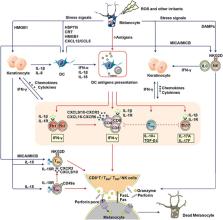
          Vitiligo is an autoimmune depigment disease results from extensive melanocytes destruction. The destruction of melanocyte is thought to be of multifactorial causation. Genome‐wide associated studies have identified single‐nucleotide polymorphisms in a panel of susceptible loci as risk factors in melanocyte death. But vitiligo onset can't be solely attributed to a susceptive genetic background. Oxidative stress triggered by elevated levels of reactive oxygen species accounts for melanocytic molecular and organelle dysfunction, a minority of melanocyte demise, and melanocyte‐specific antigens exposure. Of note, the self‐responsive immune function directly contributes to the bulk of melanocyte deaths in vitiligo. The aberrantly heightened innate immunity, type‐1‐skewed T helper, and incompetent regulatory T cells tip the balance toward autoreaction and CD8 + cytotoxic T lymphocytes finally execute the killing of melanocytes, possibly alarmed by resident memory T cells. In addition to the well‐established apoptosis and necrosis, we discuss several death modalities like oxeiptosis, ferroptosis, and necroptosis that are probably employed in melanocyte destruction. This review focuses on the various mechanisms of melanocytic death in vitiligo pathogenesis to demonstrate a panorama of that. We hope to provide new insights into vitiligo pathogenesis and treatment strategies by the review.

          Related collections

          Most cited references247

          • Record: found
          • Abstract: found
          • Article: not found

          Ferroptosis: an iron-dependent form of nonapoptotic cell death.

          Nonapoptotic forms of cell death may facilitate the selective elimination of some tumor cells or be activated in specific pathological states. The oncogenic RAS-selective lethal small molecule erastin triggers a unique iron-dependent form of nonapoptotic cell death that we term ferroptosis. Ferroptosis is dependent upon intracellular iron, but not other metals, and is morphologically, biochemically, and genetically distinct from apoptosis, necrosis, and autophagy. We identify the small molecule ferrostatin-1 as a potent inhibitor of ferroptosis in cancer cells and glutamate-induced cell death in organotypic rat brain slices, suggesting similarities between these two processes. Indeed, erastin, like glutamate, inhibits cystine uptake by the cystine/glutamate antiporter (system x(c)(-)), creating a void in the antioxidant defenses of the cell and ultimately leading to iron-dependent, oxidative death. Thus, activation of ferroptosis results in the nonapoptotic destruction of certain cancer cells, whereas inhibition of this process may protect organisms from neurodegeneration. Copyright © 2012 Elsevier Inc. All rights reserved.
            Bookmark
            • Record: found
            • Abstract: found
            • Article: not found

            Ferroptosis: A Regulated Cell Death Nexus Linking Metabolism, Redox Biology, and Disease

            Ferroptosis is a form of regulated cell death characterized by the iron-dependent accumulation of lipid hydroperoxides to lethal levels. Emerging evidence suggests that ferroptosis represents an ancient vulnerability caused by the incorporation of polyunsaturated fatty acids into cellular membranes, and cells have developed complex systems that exploit and defend against this vulnerability in different contexts. The sensitivity to ferroptosis is tightly linked to numerous biological processes, including amino acid, iron, and polyunsaturated fatty acid metabolism, and the biosynthesis of glutathione, phospholipids, NADPH, and coenzyme Q10. Ferroptosis has been implicated in the pathological cell death associated with degenerative diseases (i.e., Alzheimer's, Huntington's, and Parkinson's diseases), carcinogenesis, stroke, intracerebral hemorrhage, traumatic brain injury, ischemia-reperfusion injury, and kidney degeneration in mammals and is also implicated in heat stress in plants. Ferroptosis may also have a tumor-suppressor function that could be harnessed for cancer therapy. This Primer reviews the mechanisms underlying ferroptosis, highlights connections to other areas of biology and medicine, and recommends tools and guidelines for studying this emerging form of regulated cell death.
              Bookmark
              • Record: found
              • Abstract: found
              • Article: not found

              CD8 + T cells regulate tumor ferroptosis during cancer immunotherapy

              Summary Cancer immunotherapy restores and/or enhances effector function of CD8+ T cells in the tumor microenvironment 1,2 . CD8+ T cells activated by cancer immunotherapy execute tumor clearance mainly by inducing cell death through perforin-granzyme- and Fas/Fas ligand-pathways 3,4 . Ferroptosis is a form of cell death that differs from apoptosis and results from iron-dependent lipid peroxide accumulation 5,6 . Although it was mechanistically illuminated in vitro 7,8 , emerging evidence has shown that ferroptosis may be implicated in a variety of pathological scenarios 9,10 . However, the involvement of ferroptosis in T cell immunity and cancer immunotherapy is unknown. Here, we find that immunotherapy-activated CD8+ T cells enhance ferroptosis-specific lipid peroxidation in tumor cells, and in turn, increased ferroptosis contributes to the anti-tumor efficacy of immunotherapy. Mechanistically, interferon gamma (IFNγ) released from CD8+ T cells downregulates expression of SLC3A2 and SLC7A11, two subunits of glutamate-cystine antiporter system xc-, restrains tumor cell cystine uptake, and as a consequence, promotes tumor cell lipid peroxidation and ferroptosis. In preclinical models, depletion of cyst(e)ine by cyst(e)inase in combination with checkpoint blockade synergistically enhances T cell-mediated anti-tumor immunity and induces tumor cell ferroptosis. Expression of system xc- is negatively associated with CD8+ T cell signature, IFNγ expression, and cancer patient outcome. Transcriptome analyses before and during nivolumab therapy reveal that clinical benefits correlate with reduced expression of SLC3A2 and increased IFNγ and CD8. Thus, T cell-promoted tumor ferroptosis is a novel anti-tumor mechanism. Targeting tumor ferroptosis pathway constitutes a therapeutic approach in combination with checkpoint blockade.
                Bookmark

                Author and article information

                Contributors
                lishli@fmmu.edu.cn
                lichying@fmmu.edu.cn
                Journal
                Med Res Rev
                Med Res Rev
                10.1002/(ISSN)1098-1128
                MED
                Medicinal Research Reviews
                John Wiley and Sons Inc. (Hoboken )
                0198-6325
                1098-1128
                17 November 2020
                March 2021
                : 41
                : 2 ( doiID: 10.1002/med.v41.2 )
                : 1138-1166
                Affiliations
                [ 1 ] Department of Dermatology Xijing hospital, Fourth Military Medical University Xi'an Shannxi China
                Author notes
                [*] Correspondence Chunying Li and Shuli Li, Department of Dermatology, Xijing hospital, Fourth Military Medical University, Xi'an, 710032 Shannxi, China.

                Email: lichying@ 123456fmmu.edu.cn (C. L.) and lishli@ 123456fmmu.edu.cn (S. L.)

                Author information
                http://orcid.org/0000-0001-9369-5128
                https://orcid.org/0000-0002-2335-507X
                http://orcid.org/0000-0003-3004-5376
                Article
                MED21754
                10.1002/med.21754
                7983894
                33200838
                9deb041c-b50e-4b32-8ed2-c7fe2e6b59c0
                © 2020 The Authors. Medicinal Research Reviews published by Wiley Periodicals LLC

                This is an open access article under the terms of the http://creativecommons.org/licenses/by/4.0/ License, which permits use, distribution and reproduction in any medium, provided the original work is properly cited.

                History
                : 16 October 2020
                : 09 July 2020
                : 01 November 2020
                Page count
                Figures: 6, Tables: 0, Pages: 29, Words: 16172
                Funding
                Funded by: National Natural Science Foundation of China , open-funder-registry 10.13039/501100001809;
                Award ID: 81930087
                Award ID: 91742201
                Award ID: 81625020
                Categories
                Review Article
                Review Articles
                Custom metadata
                2.0
                March 2021
                Converter:WILEY_ML3GV2_TO_JATSPMC version:6.0.0 mode:remove_FC converted:22.03.2021

                autoimmunity,death,melanocyte,oxidative stress,vitiligo
                autoimmunity, death, melanocyte, oxidative stress, vitiligo

                Comments

                Comment on this article