55
views
0
recommends
+1 Recommend
0 collections
    0
    shares
      • Record: found
      • Abstract: found
      • Article: found
      Is Open Access

      Photostable fluorescent organic dots with aggregation-induced emission (AIE dots) for noninvasive long-term cell tracing

      research-article

      Read this article at

      Bookmark
          There is no author summary for this article yet. Authors can add summaries to their articles on ScienceOpen to make them more accessible to a non-specialist audience.

          Abstract

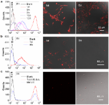
          Long-term noninvasive cell tracing by fluorescent probes is of great importance to life science and biomedical engineering. For example, understanding genesis, development, invasion and metastasis of cancerous cells and monitoring tissue regeneration after stem cell transplantation require continual tracing of the biological processes by cytocompatible fluorescent probes over a long period of time. In this work, we successfully developed organic far-red/near-infrared dots with aggregation-induced emission (AIE dots) and demonstrated their utilities as long-term cell trackers. The high emission efficiency, large absorptivity, excellent biocompatibility, and strong photobleaching resistance of the AIE dots functionalized by cell penetrating peptides derived from transactivator of transcription proteins ensured outstanding long-term noninvasive in vitro and in vivo cell tracing. The organic AIE dots outperform their counterparts of inorganic quantum dots, opening a new avenue in the development of fluorescent probes for following biological processes such as carcinogenesis.

          Related collections

          Most cited references28

          • Record: found
          • Abstract: found
          • Article: not found

          Elucidating the mechanism of cellular uptake and removal of protein-coated gold nanoparticles of different sizes and shapes.

          We investigated the mechanism by which transferrin-coated gold nanoparticles (Au NP) of different sizes and shapes entered mammalian cells. We determined that transferrin-coated Au NP entered the cells via clathrin-mediated endocytosis pathway. The NPs exocytosed out of the cells in a linear relationship to size. This was different than the relationship between uptake and size. Furthermore, we developed a mathematical equation to predict the relationship of size versus exocytosis for different cell lines. These studies will provide guidelines for developing NPs for imaging and drug delivery applications, which will require "controlling" NP accumulation rate. These studies will also have implications in determining nanotoxicity.
            Bookmark
            • Record: found
            • Abstract: found
            • Article: not found

            Water-soluble quantum dots for multiphoton fluorescence imaging in vivo.

            The use of semiconductor nanocrystals (quantum dots) as fluorescent labels for multiphoton microscopy enables multicolor imaging in demanding biological environments such as living tissue. We characterized water-soluble cadmium selenide-zinc sulfide quantum dots for multiphoton imaging in live animals. These fluorescent probes have two-photon action cross sections as high as 47,000 Goeppert-Mayer units, by far the largest of any label used in multiphoton microscopy. We visualized quantum dots dynamically through the skin of living mice, in capillaries hundreds of micrometers deep. We found no evidence of blinking (fluorescence intermittency) in solution on nanosecond to millisecond time scales.
              Bookmark
              • Record: found
              • Abstract: found
              • Article: not found

              Bioconjugated quantum dots for in vivo molecular and cellular imaging.

              Semiconductor quantum dots (QDs) are tiny light-emitting particles on the nanometer scale, and are emerging as a new class of fluorescent labels for biology and medicine. In comparison with organic dyes and fluorescent proteins, they have unique optical and electronic properties, with size-tunable light emission, superior signal brightness, resistance to photobleaching, and broad absorption spectra for simultaneous excitation of multiple fluorescence colors. QDs also provide a versatile nanoscale scaffold for designing multifunctional nanoparticles with both imaging and therapeutic functions. When linked with targeting ligands such as antibodies, peptides or small molecules, QDs can be used to target tumor biomarkers as well as tumor vasculatures with high affinity and specificity. Here we discuss the synthesis and development of state-of-the-art QD probes and their use for molecular and cellular imaging. We also examine key issues for in vivo imaging and therapy, such as nanoparticle biodistribution, pharmacokinetics, and toxicology.
                Bookmark

                Author and article information

                Journal
                Sci Rep
                Sci Rep
                Scientific Reports
                Nature Publishing Group
                2045-2322
                28 January 2013
                2013
                : 3
                : 1150
                Affiliations
                [1 ]Institute of Materials Research and Engineering , 3, Research Link, Singapore 117602
                [2 ]Department of Chemical and Biomolecular Engineering, National University of Singapore , 4 Engineering Drive 4, Singapore 117576
                [3 ]Department of Chemistry, Division of Biomedical Engineering, Institute for Advanced Study, State Key Laboratory of Molecular Neuroscience, and Institute of Molecular Functional Materials, The Hong Kong University of Science & Technology , Clear Water Bay, Kowloon, Hong Kong, China
                [4 ]SCUT-HKUST Joint Research Laboratory, Guangdong Innovative Research Team, State Key Laboratory of Luminescent Materials and Devices, South China University of Technology , Guangzhou, China, 510640
                [5 ]These authors contributed equally to this work.
                Author notes
                Article
                srep01150
                10.1038/srep01150
                3556677
                23359649
                9e69e7a6-71c2-416f-8842-1277c7a0679c
                Copyright © 2013, Macmillan Publishers Limited. All rights reserved

                This work is licensed under a Creative Commons Attribution-NonCommercial-NoDerivs 3.0 Unported License. To view a copy of this license, visit http://creativecommons.org/licenses/by-nc-nd/3.0/

                History
                : 23 October 2012
                : 11 December 2012
                Categories
                Article

                Uncategorized
                Uncategorized

                Comments

                Comment on this article