55
views
0
recommends
+1 Recommend
0 collections
    0
    shares
      • Record: found
      • Abstract: found
      • Article: not found

      Release of Inflammatory Mediators by Human Adipose Tissue Is Enhanced in Obesity and Primarily by the Nonfat Cells: A Review

      review-article
      *
      Mediators of Inflammation
      Hindawi Publishing Corporation

      Read this article at

      ScienceOpenPublisherPMC
      Bookmark
          There is no author summary for this article yet. Authors can add summaries to their articles on ScienceOpen to make them more accessible to a non-specialist audience.

          Abstract

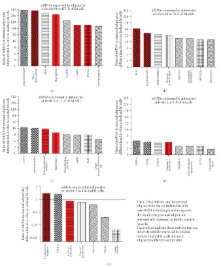
          This paper considers the role of putative adipokines that might be involved in the enhanced inflammatory response of human adipose tissue seen in obesity. Inflammatory adipokines [IL-6, IL-10, ACE, TGF β1, TNF α, IL-1 β, PAI-1, and IL-8] plus one anti-inflammatory [IL-10] adipokine were identified whose circulating levels as well as in vitro release by fat are enhanced in obesity and are primarily released by the nonfat cells of human adipose tissue. In contrast, the circulating levels of leptin and FABP-4 are also enhanced in obesity and they are primarily released by fat cells of human adipose tissue. The relative expression of adipokines and other proteins in human omental as compared to subcutaneous adipose tissue as well as their expression in the nonfat as compared to the fat cells of human omental adipose tissue is also reviewed. The conclusion is that the release of many inflammatory adipokines by adipose tissue is enhanced in obese humans.

          Related collections

          Most cited references128

          • Record: found
          • Abstract: found
          • Article: not found

          Hypoxia-inducible factor 1alpha induces fibrosis and insulin resistance in white adipose tissue.

          Adipose tissue can undergo rapid expansion during times of excess caloric intake. Like a rapidly expanding tumor mass, obese adipose tissue becomes hypoxic due to the inability of the vasculature to keep pace with tissue growth. Consequently, during the early stages of obesity, hypoxic conditions cause an increase in the level of hypoxia-inducible factor 1alpha (HIF1alpha) expression. Using a transgenic model of overexpression of a constitutively active form of HIF1alpha, we determined that HIF1alpha fails to induce the expected proangiogenic response. In contrast, we observed that HIF1alpha initiates adipose tissue fibrosis, with an associated increase in local inflammation. "Trichrome- and picrosirius red-positive streaks," enriched in fibrillar collagens, are a hallmark of adipose tissue suffering from the early stages of hypoxia-induced fibrosis. Lysyl oxidase (LOX) is a transcriptional target of HIF1alpha and acts by cross-linking collagen I and III to form the fibrillar collagen fibers. Inhibition of LOX activity by beta-aminoproprionitrile treatment results in a significant improvement in several metabolic parameters and further reduces local adipose tissue inflammation. Collectively, our observations are consistent with a model in which adipose tissue hypoxia serves as an early upstream initiator for adipose tissue dysfunction by inducing a local state of fibrosis.
            Bookmark
            • Record: found
            • Abstract: found
            • Article: not found

            Visceral and subcutaneous adipose tissue volumes are cross-sectionally related to markers of inflammation and oxidative stress: the Framingham Heart Study.

            Excess adiposity is associated with greater systemic inflammation. Whether visceral adiposity is more proinflammatory than subcutaneous abdominal adiposity is unclear. We examined the relations of abdominal subcutaneous adipose tissue (SAT) and visceral adipose tissue (VAT), assessed by multidetector computerized tomography, to circulating inflammatory and oxidative stress biomarkers in 1250 Framingham Heart Study participants (52% women; age 60+/-9 years). Biomarkers were examined in relation to increments of SAT and VAT after adjustment for age, sex, smoking, physical activity, menopause, hormone replacement therapy, alcohol, and aspirin use; additional models included body mass index and waist circumference. SAT and VAT were positively and similarly (with respect to strength of association) related to C-reactive protein, fibrinogen, intercellular adhesion molecule-1, interleukin-6, P-selectin, and tumor necrosis factor receptor-2 (multivariable model R2 0.06 to 0.28 [SAT] and 0.07 to 0.29 [VAT]). However, compared with SAT, VAT was more highly associated with urinary isoprostanes and monocyte chemoattractant protein-1 (SAT versus VAT comparison: isoprostanes, R2 0.07 versus 0.10, P=0.002; monocyte chemoattractant protein-1, R2 0.07 versus 0.08, P=0.04). When body mass index and waist circumference were added to the models, VAT remained significantly associated with only C-reactive protein (P=0.0003 for women; P=0.006 for men), interleukin-6 (P=0.01), isoprostanes (P=0.0002), and monocyte chemoattractant protein-1 (P=0.008); SAT only remained associated with fibrinogen (P=0.01). The present cross-sectional data support an association between both SAT and VAT with inflammation and oxidative stress. The data suggest that the contribution of visceral fat to inflammation may not be completely accounted for by clinical measures of obesity (body mass index and waist circumference).
              Bookmark
              • Record: found
              • Abstract: found
              • Article: not found

              Hypoxia is a potential risk factor for chronic inflammation and adiponectin reduction in adipose tissue of ob/ob and dietary obese mice.

              Chronic inflammation and reduced adiponectin are widely observed in the white adipose tissue in obesity. However, the cause of the changes remains to be identified. In this study, we provide experimental evidence that hypoxia occurs in adipose tissue in obese mice and that adipose hypoxia may contribute to the endocrine alterations. The adipose hypoxia was demonstrated by a reduction in the interstitial partial oxygen pressure (Po(2)), an increase in the hypoxia probe signal, and an elevation in expression of the hypoxia response genes in ob/ob mice. The adipose hypoxia was confirmed in dietary obese mice by expression of hypoxia response genes. In the adipose tissue, hypoxia was associated with an increased expression of inflammatory genes and decreased expression of adiponectin. In dietary obese mice, reduction in body weight by calorie restriction was associated with an improvement of oxygenation and a reduction in inflammation. In cell culture, inflammatory cytokines were induced by hypoxia in primary adipocytes and primary macrophages of lean mice. The transcription factor NF-kappaB and the TNF-alpha gene promoter were activated by hypoxia in 3T3-L1 adipocytes and NIH3T3 fibroblasts. In addition, adiponectin expression was reduced by hypoxia, and the reduction was observed in the gene promoter in adipocytes. These data suggest a potential role of hypoxia in the induction of chronic inflammation and inhibition of adiponectin in the adipose tissue in obesity.
                Bookmark

                Author and article information

                Journal
                Mediators Inflamm
                MI
                Mediators of Inflammation
                Hindawi Publishing Corporation
                0962-9351
                1466-1861
                2010
                23 May 2010
                : 2010
                : 513948
                Affiliations
                Department of Molecular Sciences, University of Tennessee Health Science Center, Memphis, TN 38163, USA
                Author notes

                Academic Editor: Giamila Fantuzzi

                Article
                10.1155/2010/513948
                2874930
                20508843
                a53855d4-3c4d-45c7-b8c5-252fbb7f8a9f
                Copyright © 2010 John N. Fain.

                This is an open access article distributed under the Creative Commons Attribution License, which permits unrestricted use, distribution, and reproduction in any medium, provided the original work is properly cited.

                History
                : 3 November 2009
                : 27 January 2010
                : 23 February 2010
                Categories
                Review Article

                Immunology
                Immunology

                Comments

                Comment on this article